. 1997 Jun 24;94(13):6904-9.
doi: 10.1073/pnas.94.13.6904.

The human cytomegalovirus US6 glycoprotein inhibits transporter associated with antigen processing-dependent peptide translocation

Affiliations

The human cytomegalovirus US6 glycoprotein inhibits transporter associated with antigen processing-dependent peptide translocation

P J Lehner et al. Proc Natl Acad Sci U S A. .

Abstract

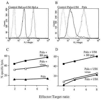
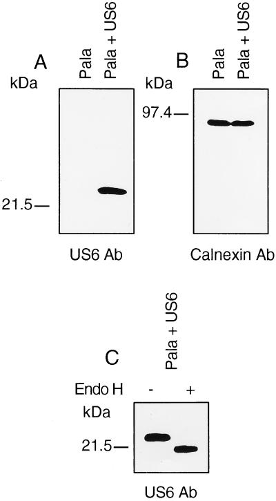
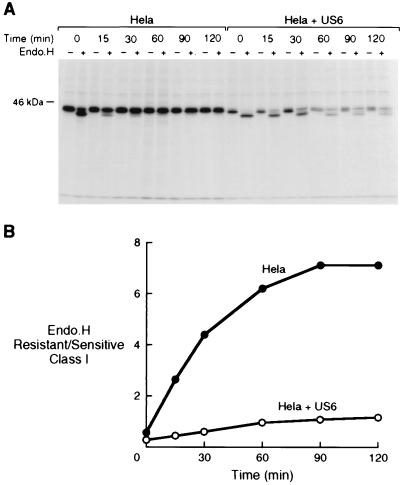
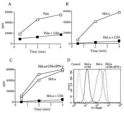
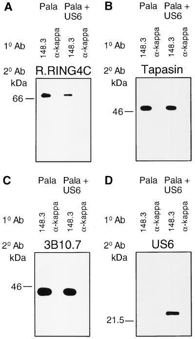
In its attempt to evade cytotoxic T cell recognition, human cytomegalovirus encodes several genes that target MHC class I molecules at different points in their assembly pathway. We show here that the human cytomegalovirus US6 gene encodes a 22-kDa glycoprotein that binds the transporter-associated with antigen processing (TAP)/class I complex and inhibits translocation of peptide from the cytosol to the endoplasmic reticulum. Major histocompatibility complex class I molecules are therefore unable to load TAP-dependent peptides, resulting in the retention of MHC class I molecules in the endoplasmic reticulum, with a consequent reduction in class I at the cell surface. Interferon-gamma treatment of US6 transfected cells overcomes this inhibition of peptide translocation and restores class I at the cell surface to wild type levels. The functional consequence of TAP inhibition is that US6 transfected cells are unable to present endogenous antigen to cytotoxic T lymphocytes and are therefore resistant to cytotoxic T lymphocyte lysis.


Figures

Figure 1
Figure 1
Expression of the US6 gene decreases cell surface expression of MHC class I molecules and prevents CTL recognition of virus-infected cells. US6 and control-transfected HeLaM cells (A) and PaLa cells (B) were analyzed for class I surface expression by flow cytometry using the mAb w6/32 and fluorescein-conjugated rabbit anti-mouse IgG. The control was a nonspecific isotype-matched monoclonal antibody. Peripheral blood mononuclear cells from an HLA-B8 positive donor were incubated in vitro with influenza A-infected cells and pulsed on day 8 with N380–88 peptide and feeder cells. On day 12 of bulk culture, CTL lysis was tested in a 51Cr release assay against Pala control (C) and Pala.US6-transfected (D) target cells, which were infected with influenza virus, pulsed with N380–88 peptide (10 μM), or untreated.
Figure 2
Figure 2
The US6 gene encodes a 22-kDa glycoprotein. US6 and control-transfected Pala cells were extracted in 1% Triton X-100 and lysates separated by SDS/12.5% PAGE, transferred to Immobilon-P membranes, and probed with anti-US6 (A) and anti-calnexin (B) antibodies. (C) Crude membrane extracts from US6 and control-transfected cells were prepared by freeze thawing. Membranes were extracted in endo H buffer, subjected to endo H or mock digestion, separated by SDS/PAGE, and after transfer to Immobilon-P membranes probed with antibody specific for US6.
Figure 3
Figure 3
Transport of class I heavy chains is inhibited in US6-transfected cells. US6 and vector control-transfected HeLaM cells were radiolabeled with [35S]methionine and [35S]cysteine for 15 min and chased for the indicated time periods. Triton X-100 lysates (1%) were immunoprecipitated with the mAb w6/32, digested or mock-digested with endo H, and subjected to 10% SDS/PAGE (A), and the ratio of endo H-resistant vs. endo H-sensitive class I heavy chains present at each time point quantitated by image analysis (B).
Figure 4
Figure 4
TAP-mediated peptide translocation is inhibited in US6-transfected cells and can be overcome with IFN-γ. Streptolysin-O (Murex)-permeabilized US6 and control-transfected Pala cells (A), HeLaM (B), and HeLaM cells that had been pretreated with IFN-γ (200 unit/ml) for 48 hr (C) were incubated with an iodinated reporter peptide at 37°C for the indicated time period, and the reaction was stopped by lysis with 3% Triton X-100, as described in Materials and Methods. Translocation into the ER was assessed by binding of the glycosylated reporter peptide to concanavalin A-Sepharose beads and counting on a γ-counter. HeLaM, US6-transfected HeLaM, and US6-transfected HeLaM cells that had been pretreated with IFN-γ were analyzed for class I surface expression by flow cytometry, using the mAb w6/32 and fluorescein-conjugated rabbit anti-mouse IgG (D). The control was a nonspecific isotype-matched monoclonal antibody.
Figure 5
Figure 5
US6 associates with the TAP/tapasin/class I complex. Digitonin lysates (1%) from US6 and control-transfected PaLa cells were immunoprecipitated with TAP1-specific (148.3) and control (κ immunoglobulin light chain-specific) mAb, coupled to A-15 m Sepharose beads. Precipitated proteins were eluted in 0.1% SDS/0.05% Triton X-100 buffer, separated by 12.5% SDS/PAGE under nonreducing conditions, transferred to Immobilon P membrane, and probed with TAP-specific, R.RING 4C (A), tapasin-specific, R.tapasinN (B), class I heavy chain-specific, 3B10.7 (C), and anti-US6 (D) antibodies.

References

    1. Reusser P, Riddell S R, Meyers J D, Greenberg P D. Blood. 1991;78:1373–1380. - PubMed
    1. Koszinowski U H, Del Val M, Reddehase M. Curr Top Microbiol Immunol. 1990;154:189–220. - PubMed
    1. Riddell S, Wantanabe K, Goodrich J, Li C R, Agha M, Greenberg P. Science. 1992;257:238–241. - PubMed
    1. Walter E A, Greenberg P D, Gilberg M J, Finch R J, Watanabe K S, Thomas E D, Riddell S R. N Engl J Med. 1995;333:1038–1044. - PubMed
    1. Beersma M F, Bijlmakers M J, Ploegh H L. J Immunol. 1993;151:4455–4464. - PubMed

Publication types

MeSH terms

Substances