. 1997 Jun 24;94(13):6927-32.
doi: 10.1073/pnas.94.13.6927.

RAB22 and RAB163/mouse BRCA2: proteins that specifically interact with the RAD51 protein

Affiliations

RAB22 and RAB163/mouse BRCA2: proteins that specifically interact with the RAD51 protein

R Mizuta et al. Proc Natl Acad Sci U S A. .

Abstract

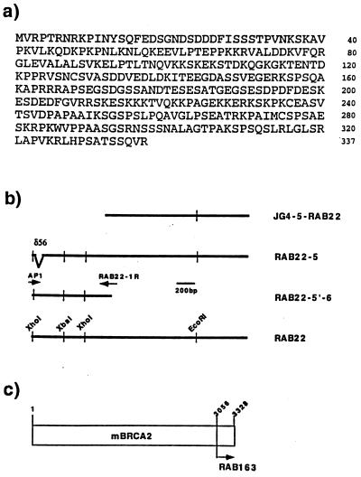
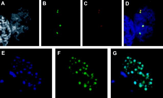
The human RAD51 protein is a homologue of the bacteria RecA and yeast RAD51 proteins that are involved in homologous recombination and DNA repair. RAD51 interacts with proteins involved in recombination and also with tumor suppressor proteins p53 and breast cancer susceptibility gene 1 (BRCA1). We have used the yeast two-hybrid system to clone murine cDNA sequences that encode two RAD51-associated molecules, RAB22 and RAB163. RAB163 encodes the C-terminal portion of mouse BRCA2, the homologue of the second breast cancer susceptibility gene protein in humans, demonstrating an in vitro association between RAD51 and BRCA2. RAB22 is a novel gene product that also interacts with RAD51 in vitro. To detect RAD51 interactions in vivo, we developed a transient nuclear focus assay that was used to demonstrate a complete colocalization of RAB22 with RAD51 in large nuclear foci.


Figures

Figure 1
Figure 1
RAD51-associated cDNA clones isolated by the yeast two-hybrid assay. (a) Predicted amino acid sequence of full-length RAB22, encoding 337 amino acids. No significant homology was observed to other sequences in GenBank. The GenBank accession number of the cDNA sequence is U93583. (b) Four representative cDNA fragments of RAB22 are shown as bold lines with the clone name listed on the right side. JG4-5–RAB22 was the original clone isolated by the yeast-two hybrid screening. RAB22-5 was cloned from a mouse thymus library using a cDNA probe prepared from JG4-5–RAB22, but contains a 56-nucleotide deletion (δ56) in the coding region. RAB22-5′-6 was fragment amplified from a mouse testis library by two primers (arrows), AP1 and RAB22-1R, and did not contain a deletion. RAB22 is the full-length cDNA clone constructed from RAB22-5 and RAB22-5′-6. The thin vertical lines are restriction enzyme sites as noted. (c) Relative position of the RAB163 peptide to the mouse BRCA2 (mBRCA2) peptide was shown by amino acid number and arrow. The full-length cDNA sequence of mBRCA2 has been reported (26).
Figure 2
Figure 2
Analysis of RAB22 expression. (Upper) Total RNA from multiple mouse tissues was probed with the full-length RAB22 cDNA. (Lower) Relative amount of total RNA in each lane is demonstrated by etidium bromide staining of 28S and 18S rRNA.
Figure 3
Figure 3
Chromosomal locations of the two RAB loci in the mouse genome. RAB loci were mapped by interspecific backcross analysis. The number of recombinant N2 animals is presented over the total number of N2 animals typed to the left of the chromsome maps between each pair of loci. The recombination frequencies, expressed as genetic distance in centimorgans (±1 SE) are also shown. The upper 95% confidence limit of the recombination distance is given in parentheses when no recombinants were found between loci. Gene order was determined by minimizing the number of recombination events required to explain the allele distribution patterns. The positions of loci on human chromosomes, where known, are shown to the right of the chromosome maps. References for the map positions of most human loci can be obtained from the Genome Data Base, a computerized database of human linkage information maintained by The William H. Welch Medical Library of The Johns Hopkins University (Baltimore).
Figure 4
Figure 4
Specific association between RAD51 and RAB22 or RAB163 in vitro by GST-affinity pull-down assay. 35S-labeled RAD51 (≈40 kDa, lane 1), RAB22 (≈45 kDa, lane 2), RAB163/mBRCA2 (≈35 kDa, lane 3), XRCC4 (≈53 kDa, lane 4), and luciferase (≈60 kDa, lane 5) were generated in vitro and visualized following SDS/PAGE. For these lanes, 2.5 μl of in vitro-translated product was separated on the gel. Each in vitro-translated protein (15 μl of reaction) also was incubated with GST–RAD51 fusion protein that was synthesized in bacteria and immobilized on glutathione-Sepharose beads (40 μl). After washing with 1× NETN buffer several times, the beads were suspended in 25 μl of 2× sample buffer (2% SDS/10% glycerol/0.01% Bromophenol Blue), boiled, and spun to pellet the beads. Then 5 μl of each supernatant was assayed by SDS/PAGE; RAD51 (lane 6), RAB22 (lane 7), RAB163/mBRCA2 (lane 8), XRCC4 (lane 9), and luciferase (lane 10).
Figure 5
Figure 5
Colocalization of RAD51 and RAB22 in discrete nuclear foci by TrNF assay. (AD) CHO-K1 cells were transiently transfected with pGFP–RAB22 and pRc/CMV–RAD51 constructs and subsequently stained with anti-RAD51 antiserum and a rhodamine-conjugated secondary antibody. A representative nucleus of a transfection-positive cell is shown. (A) DAPI nuclear counterstain in grayscale. (B) pGFP–RAB22 localization, green foci. (C) pRc/CMV–RAD51 localization by antibody detection, red foci. No foci were observed with preimmune serum (data not shown). (D) Merged image of AC. Where green and red signals overlap, a yellow signal is observed, indicating colocalization of RAB22 and RAD51. DNA counterstain is in blue. (EG) CHO-K1 cells were transiently cotransfected with pGFP–RAB22 and pBFP–RAD51 constructs and directly analyzed for nuclear foci. (E) pBFP–RAD51 localization, blue foci. (F) pGFP–RAB22 localization, green foci. (G) In merged image of E and F, aqua foci indicate colocalization of blue and green signals.

References

    1. Shinohara A, Ogawa H, Ogawa T. Cell. 1992;69:457–470. - PubMed
    1. Edelmann W, Kucherlapati R. Proc Natl Acad Sci USA. 1996;93:6225–6227. - PMC - PubMed
    1. Terasawa M, Shinohara A, Hotta Y, Ogawa H, Ogawa T. Genes Dev. 1995;9:925–934. - PubMed
    1. Sung P, Robberson D L. Cell. 1995;82:453–461. - PubMed
    1. Baumann P, Benson F E, West S C. Cell. 1996;87:757–766. - PubMed

Publication types

MeSH terms

Associated data