. 1997 Jun 24;94(13):6933-8.
doi: 10.1073/pnas.94.13.6933.

Loss of the retinoblastoma protein-related p130 protein in small cell lung carcinoma

Affiliations

Loss of the retinoblastoma protein-related p130 protein in small cell lung carcinoma

K Helin et al. Proc Natl Acad Sci U S A. .

Abstract

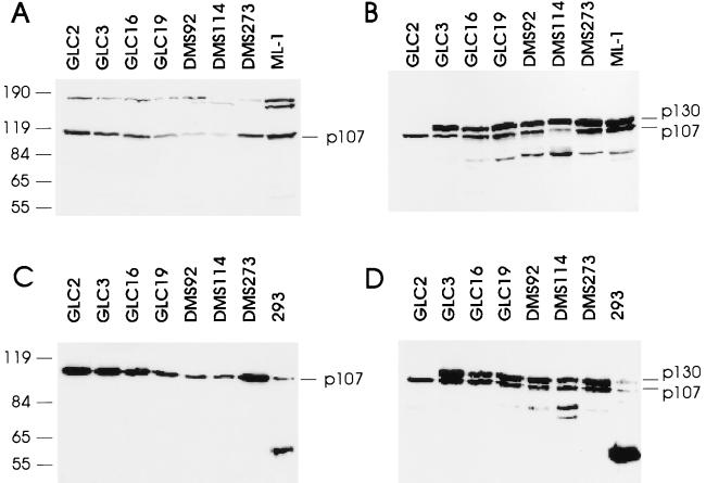
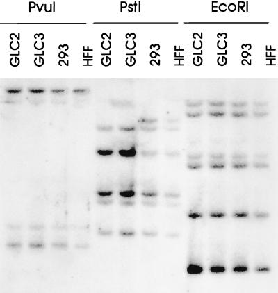
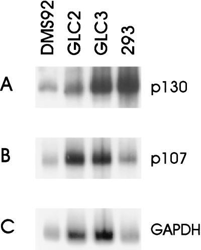
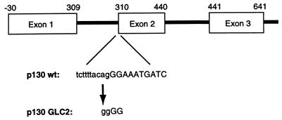
The retinoblastoma gene family consists of the tumor suppressor protein pRB and its two relatives p107 and p130. These proteins have been implicated in the regulation of cell cycle progression, in part, through inactivation of members of the E2F transcription factor family. Overexpression of pRB, p107, or p130 leads to growth arrest in the G1 phase of the cell cycle, and this arrest is abolished by complex formation with the adenovirus E1A, human papilloma virus E7, or simian virus 40 T oncoproteins. Inactivation of pRB by gross structural alterations or point mutations in the RB-1 gene has been described in a variety of human tumors, including retinoblastomas, osteosarcomas, and small cell lung carcinomas. Despite the structural and functional similarity between pRB, p107, and p130, alterations in the latter two proteins have not been identified in human tumors. We have screened a panel of 17 small cell lung carcinoma cell lines for the presence of functional p107 and p130 by evaluating their ability to form complexes with E1A in vitro. In the GLC2 small cell lung carcinoma cells no p130 protein was detected. The loss of the p130 protein is the result of a single point mutation within a splice acceptor sequence in the GLC2 genomic DNA. This mutation eliminates exon 2, leading to an in-frame stop codon, and no detectable protein is produced. These data are, to our knowledge, the first to describe the loss of p130 as a consequence of a genetic alteration, suggesting that not only pRB but also the other members of the family may contribute to tumorigenesis, providing a rationale for the observation that the DNA tumor viruses selectively target all the members of the retinoblastoma protein family.


Figures

Figure 1
Figure 1
Loss of p130 expression in the GLC2 cell line. (A) Fifty micrograms of cell lysate from the indicated cell lines was separated on an SDS/8% polyacrylamide gel and processed for Western blotting. The ML-1 cell line is a control for the migration of wild-type p107. This blot was probed with a polyclonal rabbit antibody raised against p107. (B) Fifty micrograms of cell lysate was processed as described above. The blot was probed with a monoclonal antibody to p130, Z83, that also recognizes p107, and two related proteins (or breakdown products) of 80 and 90 kDa. (C) Five hundred micrograms of cell lysate was incubated with 5 μg of GSTE1A-(1–139) for 2 h on ice, and GSTE1A-(1–139) and associated proteins were precipitated and processed for Western blotting. E1A and associated proteins from 293 cells precipitated with the monoclonal antibody M73 served as a control for the migration of p107 and p130. The blot was probed with the rabbit polyclonal antibody to p107. (D) As in C; however, the blot was probed with Z83. At the left are positions of molecular mass markers, indicated in kDa.
Figure 2
Figure 2
The structural organization of the p130 gene appears normal in GLC2 cells. Ten micrograms of genomic DNA isolated from the indicated cell lines was digested with PvuI, PstI, or EcoRI, and fragments were separated on an 0.8% agarose gel and processed for Southern blotting. The blot was probed with 32P-labeled full-length p130 cDNA.
Figure 3
Figure 3
Normal-sized mRNA for p130 is present in GLC2 cells. The RNA from the indicated cell lines was processed for Northern blotting, and the blots were probed with p130 cDNA (A), p107 cDNA (B), or glyceraldehyde-3-phosphate dehydrogenase (GAPDH) cDNA (C).
Figure 4
Figure 4
Exon–intron structure of the p130 gene with the indicated splice acceptor mutation in the p130 gene present in GLC2 cells. Numbers above the exons indicate the nucleotide numbers in the wild-type p130 cDNA.
Figure 5
Figure 5
FISH analysis of cell line GLC2. (A) Chromosome 16 paint showing the normal (n), and the two rearranged 16s (q+, p+) observed in 75% of the cells. (B) FISH signals (arrows) at 16q12–13 on all three chromosomes 16 obtained with the p130 gene. (C) Chromosome 16 paint showing the two normal (n) and the single rearranged (q+) chromosome 16 observed in 25% of the cells. (D) FISH signals (arrows) of p130 at 16q12–13 on all three chromosomes 16 obtained with the p130 gene.

References

    1. Weinberg R A. Cell. 1995;81:323–330. - PubMed
    1. Scheffner M, Münger K, Byrne J C, Howley P M. Proc Natl Acad Sci USA. 1991;88:5523–5527. - PMC - PubMed
    1. Vousden K H. Semin Cancer Biol. 1995;6:109–116. - PubMed
    1. Cobrinik D. Curr Top Microbiol Immunol. 1996;208:31–60. - PubMed
    1. Adams P D, Kaelin W G., Jr Curr Top Microbiol Immunol. 1996;208:79–93. - PubMed

Publication types

MeSH terms