. 1997 Jun 24;94(13):6948-53.
doi: 10.1073/pnas.94.13.6948.

Molecular delineation of the smallest commonly deleted region of chromosome 5 in malignant myeloid diseases to 1-1.5 Mb and preparation of a PAC-based physical map

Affiliations

Molecular delineation of the smallest commonly deleted region of chromosome 5 in malignant myeloid diseases to 1-1.5 Mb and preparation of a PAC-based physical map

N Zhao et al. Proc Natl Acad Sci U S A. .

Abstract

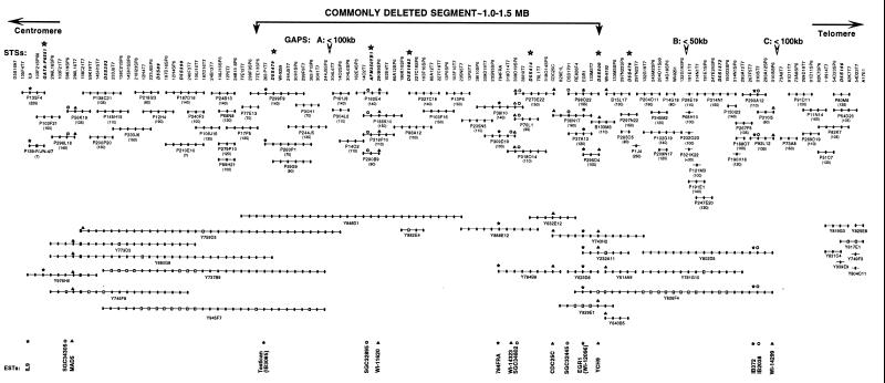
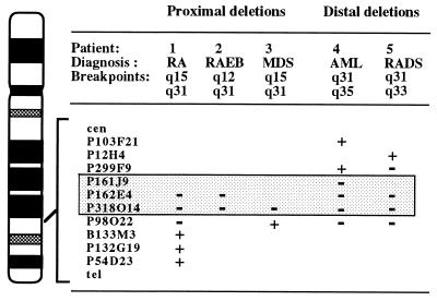
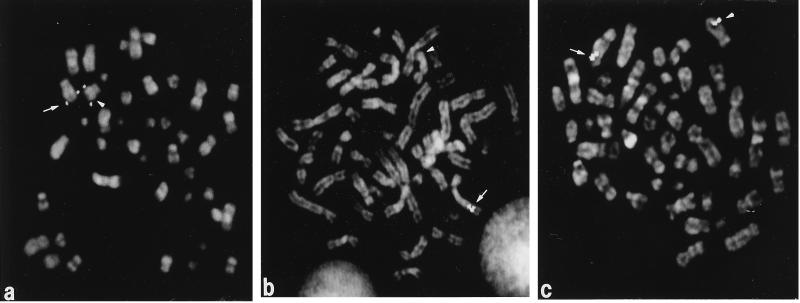
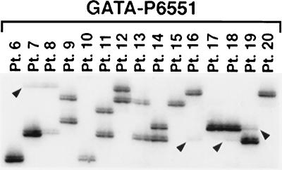
Loss of a whole chromosome 5 or a deletion of the long arm, del(5q), is a recurring abnormality in malignant myeloid diseases. In previous studies, we delineated a commonly deleted segment of approximately 4 Mb within band 5q31 that was flanked by IL9 on the proximal side and D5S166 on the distal side. We have generated a physical map of P1 (PAC), bacterial (BAC), and yeast artificial chromosome (YAC) clones of this interval. The contig consists of 108 clones (78 PACs, 2 BACs, and 28 YACs) to which 125 markers (5 genes, 11 expressed sequence tags, 12 polymorphisms, and 97 sequence-tagged sites) have been mapped. Using PAC clones for fluorescence in situ hybridization analysis of leukemia cells with a del(5q), we have narrowed the commonly deleted segment to 1-1.5 Mb between D5S479 and D5S500. To search for allele loss, we used 7 microsatellite markers within and flanking the commonly deleted segment to examine leukemia cells from 28 patients with loss of 5q, and 14 patients without cytogenetically detectable loss of 5q. In the first group of patients, we detected hemizygous deletions, consistent with the cytogenetically visible loss; no homozygous deletions were detected. No allele loss was detected in patients without abnormalities of chromosome 5, suggesting that allele loss on 5q is the result of visible chromosomal abnormalities. The development of a stable PAC contig and the identification of the smallest commonly deleted segment will facilitate the molecular cloning of a myeloid leukemia suppressor gene on 5q.


Figures

Figure 1
Figure 1
STS content map of the genomic contig of 5q31. PAC, BAC, and YAC clones are prefaced by a P, B, or Y, respectively. The clones are presented as lines; their size reflects the number of STSs contained in each clone. Physical size in kilobases is given in braces. Solid circles indicate positive STSs. Open squares indicate that a clone was found to be negative or ambiguous for a given STS. STS markers are listed at the top of the map; those indicated by an asterisk represent microsatellite markers used for allele loss studies in leukemia cells. Gaps in the contig are indicated by an open arrow at the top of the map, and the estimated sizes of the gaps in kilobases are listed. The location of known genes and ESTs are shown at the bottom of the map.
Figure 2
Figure 2
Diagram of the banding pattern of chromosome 5 illustrating the results of FISH analysis of five patients with a del(5q) involving either a proximal (two patients) or distal (three patients) breakpoint in 5q31. The PAC clones are shown on the right of the chromosome. Hybridization results for the deleted homologs are indicated by a “+” (positive signal) or “−” (no signal) sign. The genomic segment between P299F9 and P98O22 (shaded box) was deleted in each patient examined. RA, refractory anemia; RAEB, RA with excess blasts; RADS, RA with dysplasia. Patients 1 and 4 were also examined in a previous report (patients 5 and 9, respectively, in ref. 9).
Figure 3
Figure 3
PAC clones within the contig were hybridized to metaphase cells from patients with a del(5q). (a) PAC P299F9 hybridized to both the normal (arrow) and deleted (arrowhead) homologs in leukemia cells with a del(5)(q31q35) (patient 4 in Fig. 2). Thus, it is proximal to the commonly deleted segment. (b) Clone P318O14 is deleted on the rearranged homolog (arrowhead) in leukemia cells with a del(5)(q15q31) (patient 1 in Fig. 2). (c) Clone P98O22 is retained on the deleted (arrowhead) and normal (arrow) homologs in leukemia cells with a del(5)(q15q31) (patient 3 in Fig. 2), and is distal to the commonly deleted segment.
Figure 4
Figure 4
Allele-loss studies of DNA from myeloid leukemia cells using the GATA-P6551 (D5S816) STRP centromeric to the commonly deleted segment. Hemizygous loss of alleles is indicated with arrowheads. Patients 6–8, 10, 13, and 15–20 had cytogenetic abnormalities leading to loss of 5q, whereas patients 9, 11, 12, and 14 had no visible loss of chromosomal material from 5q. Single bands are visible for patients 6, 10, 15, 17, and 20; thus, we cannot distinguish between homozygosity and complete loss of one allele.

References

    1. Le Beau M M, Rowley J D. In: Hematology. Beutler E, Erslev A J, Lichtman M A, Coller B S, Kipps T J, editors. New York: McGraw–Hill; 1995. pp. 98–106.
    1. Mitelman F. Catalog of Chromosome Aberrations in Cancer. 5th Ed. New York: Wiley; 1994.
    1. Mitelman F, Kaneko Y, Berger R. In: Human Gene Mapping 1993. Cuticchia A J, Pearson P L, editors. Baltimore, MD: Johns Hopkins Univ. Press; 1994. pp. 773–812.
    1. Rabbitts T H. Nature (London) 1994;372:143–149. - PubMed
    1. Knudson A G. Proc Natl Acad Sci USA. 1993;90:10914–10921. - PMC - PubMed

Publication types

LinkOut - more resources