. 1997 Jun 24;94(13):6954-8.
doi: 10.1073/pnas.94.13.6954.

Superoxide and peroxynitrite generation from inducible nitric oxide synthase in macrophages

Affiliations

Superoxide and peroxynitrite generation from inducible nitric oxide synthase in macrophages

Y Xia et al. Proc Natl Acad Sci U S A. .

Abstract

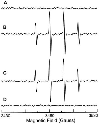
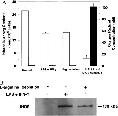
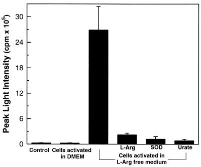
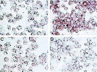
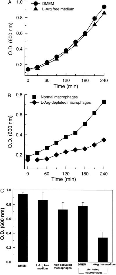
Superoxide (O-2) and nitric oxide (NO) act to kill invading microbes in phagocytes. In macrophages NO is synthesized by inducible nitric oxide synthase (iNOS, NOS 2) from L-arginine (L-Arg) and oxygen; however, O-2 was thought to be produced mainly by NADPH oxidase. Electron paramagnetic resonance (EPR) spin trapping experiments performed in murine macrophages demonstrate a novel pathway of O-2 generation. It was observed that depletion of cytosolic L-Arg triggers O-2 generation from iNOS. This iNOS-mediated O-2 generation was blocked by the NOS inhibitor N-nitro-L-arginine methyl ester or by L-Arg, but not by the noninhibitory enantiomer N-nitro-D-arginine methyl ester. In L-Arg-depleted macrophages iNOS generates both O-2 and NO that interact to form the potent oxidant peroxynitrite (ONOO-), which was detected by luminol luminescence and whose formation was blocked by superoxide dismutase, urate, or L-Arg. This iNOS-derived ONOO- resulted in nitrotyrosine formation, and this was inhibited by iNOS blockade. iNOS-mediated O-2 and ONOO- increased the antibacterial activity of macrophages. Thus, with reduced L-Arg availability iNOS produces O-2 and ONOO- that modulate macrophage function. Due to the existence of L-Arg depletion in inflammation, iNOS-mediated O-2 and ONOO- may occur and contribute to cytostatic/cytotoxic actions of macrophages.


Figures

Figure 1
Figure 1
iNOS-mediated oxygen radical generation in macrophages measured by EPR spin trapping. EPR spectra were obtained in the presence of 50 mM DMPO from the following preparations. Trace A, normal cultured cells. Trace B, cells after 24-hr activation with LPS and IFN-γ in DMEM. Trace C, cells incubated in l-Arg-free medium for 24 hr. Trace D, cells after 24-hr activation with LPS and IFN-γ in l-Arg-free medium (l-Arg-depleted cells). Trace E, l-Arg-depleted cells with SOD (200 units/ml). Trace F, l-Arg-depleted cells with 1 mM l-NAME. Trace G, l-Arg-depleted cells with 1 mM d-NAME. Trace H, l-Arg-depleted cells with 1 mM l-NMMA. Trace I, l-Arg-depleted cells with catalase (300 units/ml). Representative spectra were shown from triplicate measurements.
Figure 2
Figure 2
Effects of l-NAME or iNOS induction on NADPH oxidase-mediated oxygen radical generation in macrophages. EPR spin trapping was performed in the presence of 50 mM DMPO. Trace A, control macrophages. Trace B, after activation of NADPH oxidase with 200 ng/ml PMA. Trace C, activation of NADPH oxidase with 200 ng/ml PMA in the presence of 1 mM l-NAME. Trace D, macrophages prestimulated for 24 hr with LPS and IFN-γ with addition of 200 ng/ml PMA. The NOS inhibitor l-NAME did not alter NADPH oxidase-mediated radical generation, but prestimulation with LPS and IFN-γ totally blocked this radical generation.
Figure 3
Figure 3
Effects of l-Arg on oxygen radical formation and iNOS expression. (A) Relationship between intracellular l-Arg levels (open bars) and oxygen radical production (filled bars) in macrophages. Control, normal cultured cells; LPS + IFN-γ, cells after 24-hr activation in DMEM; l-Arg depletion, cells incubated in l-Arg-free medium for 24 hr; LPS + IFN-γ and l-Arg depletion, cells after 24-hr activation in l-Arg-free medium. Data are expressed as mean ± SEM obtained from three experiments. (B) Western blot analysis of iNOS protein in nonactivated cells and cells activated in the presence or absence of l-Arg. While no iNOS could be detected in nonactivated cells, identical amounts of iNOS protein were observed in cells activated by LPS and IFN-γ in either normal DMEM or l-Arg-free medium.
Figure 4
Figure 4
iNOS-mediated ONOO generation in l-Arg-depleted macrophages. Although no luminescence was detected in control cells or cells activated in normal DMEM, strong luminescence was seen in l-Arg-depleted macrophages. This luminescence was blocked by l-Arg (1 mM), SOD (200 units/ml), or urate (1 mM).
Figure 5
Figure 5
Nitrotyrosine formation in l-Arg-depleted macrophages. Prominent immunostaining of nitrotyrosine was observed in the cells activated in l-Arg-free medium (B) but not in the cells activated in normal DMEM (A). This staining was blocked by 1 mM l-NAME (C) and preincubation of the primary antibody with 1 mM nitrotyrosine (D). (×400.)
Figure 6
Figure 6
Inhibitory effects of iNOS-mediated O2⨪ and ONOO on the growth of E. coli. (A) Growth of E. coli in DMEM and l-Arg-free medium. (B) Growth of E. coli in the presence of normal RAW 264.7 cells and cells preactivated in l-Arg-free medium (l-Arg-depleted macrophages). Results are the average of three experiments. (C) E. coli levels in the medium after 4-hr incubations.

References

    1. Bastian N R, Hibbs J B., Jr Curr Opin Immunol. 1994;6:131–139. - PubMed
    1. Nathan C, Xie Q-w. J Biol Chem. 1994;269:13725–13728. - PubMed
    1. Rosen G M, Pou S, Ramos C L, Cohen M S, Britigan B E. FASEB J. 1995;9:200–209. - PubMed
    1. Nathan C, Xie Q-w. Cell. 1994;78:915–918. - PubMed
    1. Pou S, Pou W S, Bredt D S, Snyder S H, Rosen G M. J Biol Chem. 1992;267:24173–24176. - PubMed

Publication types

LinkOut - more resources