. 1997 Jun 24;94(13):7018-23.
doi: 10.1073/pnas.94.13.7018.

Neurturin shares receptors and signal transduction pathways with glial cell line-derived neurotrophic factor in sympathetic neurons

Affiliations

Neurturin shares receptors and signal transduction pathways with glial cell line-derived neurotrophic factor in sympathetic neurons

D J Creedon et al. Proc Natl Acad Sci U S A. .

Abstract

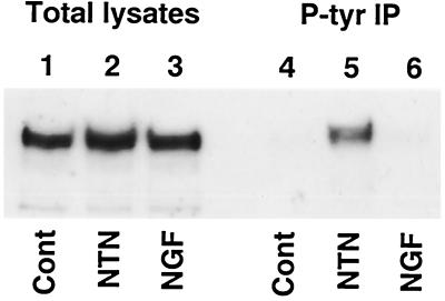
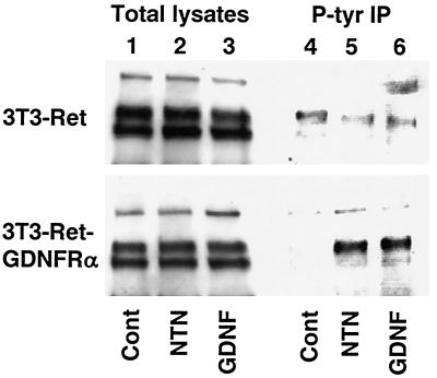
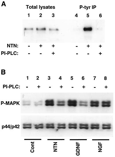
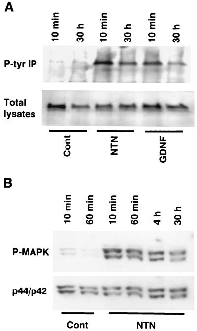
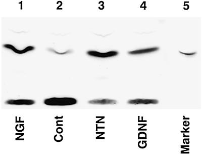
Neurturin (NTN) is a neurotrophic factor that shares homology with glial cell line-derived neurotrophic factor (GDNF). Recently, a receptor complex has been identified for GDNF that includes the Ret tyrosine kinase receptor and a glycosylphosphatidylinositol-linked protein termed "GDNFRalpha." However, differences in the phenotype of Ret and GDNF knockout animals suggest that Ret has at least one additional ligand. In this report, we demonstrate that NTN induces Ret phosphorylation in primary cultures of rat superior cervical ganglion (SCG) neurons. NTN also caused Ret phosphorylation in fibroblasts that were transfected stably with Ret and GDNFRalpha but not in cells expressing Ret alone. A glycosylphosphatidylinositol-linked protein also was important for NTN and GDNF signaling in SCG neurons; phosphatidylinositol-specific phospholipase C treatment of SCG cultures reduced the ability of NTN to phosphorylate Ret and the ability of NTN or GDNF to activate the mitogen-activated protein kinase pathway. NTN and GDNF also caused sustained activation of Ret and the mitogen-activated protein kinase pathway in SCG neurons. Finally, both NTN and GDNF activated the phosphatidylinositol 3-kinase pathway in SCG neurons, which may be important for the ability of NTN and GDNF to promote neuronal survival. These data indicate that NTN is a physiologically relevant ligand for the Ret receptor and suggest that NTN may have a critical role in the development of many neuronal populations.


Figures

Figure 1
Figure 1
NTN induces Ret tyrosine-phosphorylation in SCG neurons. SCG cultures were deprived of NGF for 4 h then treated with medium containing no additions [Control (Cont), lanes 1 and 4], 50 ng/ml NTN (lanes 2 and 5), or 50 ng/ml NGF (lanes 3 and 6). After 10 min, the cultures were lysed. Protein immunoblots were probed with an anti-Ret antibody; lanes 1–3 contain total lysates, and lanes 4–6 contain phosphotyrosine immunoprecipitates of the corresponding samples.
Figure 2
Figure 2
NTN induces Ret phosphorylation in fibroblasts expressing Ret and GDNFRα but not in those expressing Ret alone. NIH 3T3 fibroblasts expressing Ret alone (Upper) or Ret and GDNFRα (Lower) were treated with medium containing no additions [Control (Cont), lanes 1 and 4], 50 ng/ml NTN (lanes 2 and 5), or 50 ng/ml GDNF (lanes 3 and 6). Western blots of total protein (lanes 1–3) or phosphotyrosine immunoprecipitates (lanes 4–6) were probed with an anti-Ret antibody.
Figure 3
Figure 3
Cleavage of GPI linkages reduces the ability of NTN to induce Ret phosphorylation or MAPK activation. (A) Before the addition of 20 ng/ml NTN for 10 min, SCG cultures were treated for 2 h with or without PI-PLC to cleave GPI-linked proteins from the cell membrane. Western blots of total lysates (lanes 1–3) and corresponding phosphotyrosine immunoprecipitates (lanes 4–6) were probed with an anti-Ret antibody. (B) SCG neurons treated with or without PI-PLC for 2 h were then treated for 10 min with medium containing no additions (lanes 1 and 2), 20 ng/ml NTN (lanes 3 and 4), 20 ng/ml GDNF (lanes 5 and 6), or 20 ng/ml NGF (lanes 7 and 8). A protein blot of total cell lysates was first probed with a phospho-specific MAPK antibody (P-MAPK; Upper); the same blot was then re-probed with an antibody that recognizes phosphorylated and nonphosphorylated MAPK (p44/p42; Lower).
Figure 4
Figure 4
NTN causes sustained activation of Ret and MAPK. (A) SCG cultures were treated with control medium (Cont), NTN, or GDNF and lysed either 10 min or 30 h after treatment. Western blots of phosphotyrosine immunoprecipitates (Upper) and corresponding total lysates (Lower) were probed with an anti-Ret antibody. (B) SCG cultures were deprived of NGF for 2 h then treated with control medium (Cont) or NTN for 10 min, 60 min, 4 h, or 30 h. A protein blot of total cell lysates was first probed with a phospho-specific MAPK antibody (P-MAPK; Upper) and then re-probed with an antibody that recognizes phosphorylated and nonphosphorylated MAPK (p44/p42; Lower).
Figure 5
Figure 5
NTN and GDNF activate PI-3-K. SCG cultures were deprived of NGF for 4 h and then treated with medium containing 50 ng/ml NGF (lane 1), control medium (Cont; lane 2), 50 ng/ml NTN (lane 3), or 50 ng/ml GDNF (lane 4) for 30 min. The cultures were lysed, and phosphotyrosine immunoprecipitates were assayed for PI-3-K activity. Lane 5 contains a 32P-radiolabeled phosphatidylinositol-3-phosphate marker.

References

    1. Kotzbauer P T, Lampe P A, Heuckeroth R O, Golden J P, Creedon D J, Johnson E M, Jr, Milbrandt J. Nature. 1996;384:467–470. - PubMed
    1. Trupp M, Arenas E, Fainzilber M, Nilsson A S, Sieber B A, Grigoriou M, Kilkenny C, Salazar-Grueso E, Pachnis V, Arumäe U, Sariola H, Saarma M, Ibáñez C F. Nature (London) 1996;381:785–788. - PubMed
    1. Treanor J J, Goodman L, de Sauvage F, Stone D M, Poulsen K T, et al. Nature (London) 1996;382:80–83. - PubMed
    1. Jing S, Wen D, Yu Y, Holst P L, Luo Y, Fang M, Tamir R, Antonio L, Hu Z, Cupples R, Louis J C, Hu S, Altrock B W, Fox G M. Cell. 1996;85:1113–1124. - PubMed
    1. Durbec P, Marcos-Gutierrez C V, Kilkenny C, Grigoriou M, Wartiowaara K, Suvanto P, Smith D, Ponder B, Costantini F, Saarma M, Sariola H, Pachnis V. Nature (London) 1996;381:789–793. - PubMed

Publication types

MeSH terms

LinkOut - more resources