. 1997 Jun 24;94(13):7024-9.
doi: 10.1073/pnas.94.13.7024.

Glutamine synthetase protects against neuronal degeneration in injured retinal tissue

Affiliations

Glutamine synthetase protects against neuronal degeneration in injured retinal tissue

R Gorovits et al. Proc Natl Acad Sci U S A. .

Abstract

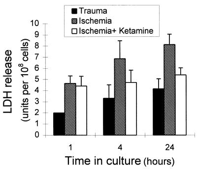
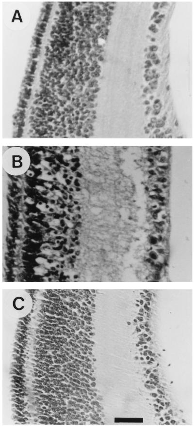
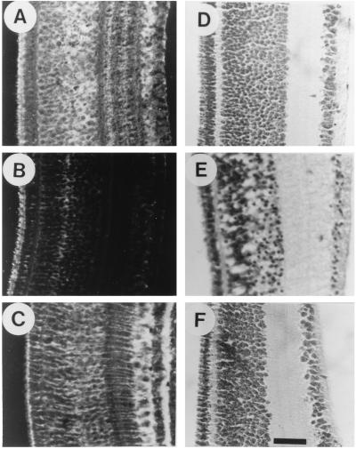
The neurotransmitter glutamate is neurotoxic when it is accumulated in a massive amount in the extracellular fluid. Excessive release of glutamate has been shown to be a major cause of neuronal degeneration after central nervous system injury. Under normal conditions, accumulation of synaptically released glutamate is prevented, at least in part, by a glial uptake system in which the glia-specific enzyme glutamine synthetase (GS) plays a key role. We postulated that glial cells cannot cope with glutamate neurotoxicity because the level of GS is not high enough to catalyze the excessive amounts of glutamate released by damaged neurons. We examined whether elevation of GS expression in glial cells protects against neuronal degeneration in injured retinal tissue. Analysis of lactate dehydrogenase efflux, DNA fragmentation, and histological sections revealed that hormonal induction of the endogenous GS gene in retinal glial cells correlates with a decline in neuronal degeneration, whereas inhibition of GS activity by methionine sulfoximine leads to increased cell death. A supply of purified GS enzyme to the culture medium of retinal explants or directly to the embryo in ovo causes a dose-dependent decline in the extent of cell death. These results show that GS is a potent neuroprotectant and that elevation of GS expression in glial cells activates an endogenous mechanism whereby neurons are protected from the deleterious effects of excess glutamate in extracellular fluid after trauma or ischemia. Our results suggest new approaches to the clinical handling of neuronal degeneration.


Figures

Figure 1
Figure 1
LDH release by retinal tissue exposed to trauma or ischemia. Retinal tissue from E17 embryos was exposed to trauma by the process of excising the tissue and subsequently cutting it into pieces. The tissue was organ-cultured for 24 h, and LDH release was measured in samples of the culture medium 1, 4, and 24 h after tissue excision (solid bars). E17 retina was exposed to ischemia by organ-culturing the tissue pieces for 50 min in glucose-free medium in flasks that were gassed with 95% N2/5% CO2 (hatched bars). In some experiments the culture medium of the ischemic tissue contained the glutamate antagonist ketamine (10 mM/ml) (open bars). The levels of LDH were measured in medium samples 1, 4, and 24 h after tissue excision. Each bar represents the mean ± SD of three separate experiments, each performed in triplicate (n = 9).
Figure 2
Figure 2
Inverse correlation between GS induction and LDH release. (A) Cortisol (1 mg per egg) was injected into E15, E16, or E17 eggs, which were then incubated for an additional 48, 24, or 4 h, respectively. Retinal tissue was excised, cut into pieces, and organ-cultured for 4 h. The levels of GS in the tissue and of LDH in the culture medium were measured. (B) E15 eggs were injected with cortisol in the indicated amounts and incubated for an additional 48 h. Retinal tissues were excised, cut into pieces, and organ-cultured for 4 h. The level of GS in the tissue and of LDH in the culture medium was measured. (C) E15 eggs were coinjected with cortisol (1 mg per egg) and MSO (1 mg per egg), buthionine sulfoximine (BSO; 1 mg per egg), or carrier. The eggs were incubated for an additional 48 h before the retina was excised. Retinal explants were cut into pieces and organ-cultured for 4 h. The levels of GS in the tissue and of LDH in the culture medium were measured. The levels of GS and LDH obtain after coinjection of cortisol and carrier were given the arbitrary value of 100. Results are means ± SD of three independent experiments, each performed in duplicate (n = 6).
Figure 3
Figure 3
Morphological evidence for cortisol protection against neuronal degeneration. E15 eggs were injected with cortisol (1 mg per egg) (A) or carrier (B) and incubated for an additional 48 h before the retina was excised. Retinal explants were cut into pieces, organ-cultured for 4 h, and then embedded in paraffin. Untreated E17 retina was embedded immediately after excision (C). Paraffin sections were stained with hematoxylin/eosin. (Bar = 20 μm.)
Figure 4
Figure 4
Internucleosomal DNA fragmentation is attenuated by cortisol. E15 eggs were injected with cortisol (1 mg per egg; lane 4) or carrier (lane 3) and incubated for an additional 48 h before the retina was excised. Retinal explants were cut into pieces and organ-cultured for 24 h. DNA was extracted, and samples from an equal numbers of cells (5 × 10⁷ cells per lane) were analyzed by gel electrophorsis and visualized with the fluorescent intercalating dye ethidium bromide. DNA was extracted from untreated E17 retina immediately after tissue excision (lane 2). Molecular weight markers are from a HaeIII digest of ØX174 phage DNA (lane 1).
Figure 5
Figure 5
Supply of purified GS enzyme in vitro or in ovo reduces the extent of LDH release. (A) E17 retina was excised, cut into pieces, and organ-cultured in the absence (−) or presence of purified GS enzyme in the indicated unit amounts. The level of LDH in the culture medium was measured after 4 h. Each bar represents the mean ± SD of two separate experiments, each performed in triplicate (n = 6). (B) E17 eggs were injected with carrier (−), with purified GS enzyme in the indicated unit amounts (1 to 8 units per egg), with ovalbumin (20 μg per egg; OVA), or with heat-inactivated GS enzyme (2 units per egg; IGS). The injected eggs were incubated for 2 h before the retina was excised. Retinal explants were cut into pieces and organ-cultured for 4 h, and the level of LDH in the culture medium was then measured. Each bar represents the mean ± SD of two separate experiments, each performed in quadruplicate (n = 8). (C) E15, E16, and E17 eggs were injected with purified GS enzyme (2 units per egg). Retinas were excised from the injected E17 eggs immediately (−) or after 2 or 4 h of incubation. Injected E16 and E15 eggs were incubated for an additional 24 or 48 h, respectively, before the retinal tissues were excised. Tissue explants were cut into pieces and organ-cultured for 4 h, and the level of LDH in the culture medium was then measured. Each bar represents the mean ± SD of two separate experiments, each performed in quadruplicate (n = 8).
Figure 6
Figure 6
Morphological evidence for protective effects of the purified GS enzyme. E17 eggs were injected with purified GS enzyme (2 units per egg; A and D) or with carrier (B and E) and incubated for 2 h before the retinas were excised. Retinal explants were embedded immediately after excision (A and B) or were cut into pieces, organ-cultured for 4 h, and then embedded in paraffin (D and E). Paraffin sections were stained with rabbit anti-GS-specific antiserum and with goat anti-rabbit IgG (A and B) or with hematoxylin/eosin (D and E). E15 eggs were injected with cortisol (1 mg per egg) and incubated for an additional 48 h before the retina was excised. The excised tissue was embedded in paraffin, and histological sections were stained with rabbit anti-GS-specific antiserum and with goat anti-rabbit IgG (C). Untreated E17 retina was embedded immediately after excision and stained with hematoxylin/eosin (F). (Bar = 25 μm.)

Similar articles

Cited by

References

    1. Rothman S M, Olney J W. Ann Neurol. 1986;19:105–111. - PubMed
    1. Choi D W, Rothman S M. Annu Rev Neurosci. 1990;13:171–182. - PubMed
    1. Van der Berg C J, Garfinkel D. Biochem J. 1971;123:211–218. - PMC - PubMed
    1. Kennedy A J, Voaden M J, Marshall J. Nature (London) 1974;252:50–52. - PubMed
    1. Moscona A A. In: Progress in Retinal Research. Osborne N N, Chader G J, editors. Vol. 2. Oxford: Permagon; 1983. pp. 111–135.

Publication types

Substances

LinkOut - more resources