. 1997 Jun 24;94(13):7070-5.
doi: 10.1073/pnas.94.13.7070.

Activation of heteromeric G protein-gated inward rectifier K+ channels overexpressed by adenovirus gene transfer inhibits the excitability of hippocampal neurons

Affiliations

Activation of heteromeric G protein-gated inward rectifier K+ channels overexpressed by adenovirus gene transfer inhibits the excitability of hippocampal neurons

M U Ehrengruber et al. Proc Natl Acad Sci U S A. .

Erratum in

  • Proc Natl Acad Sci U S A 1997 Aug 19;94(17):9511

Abstract

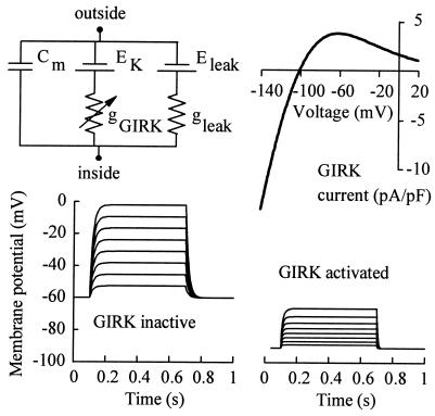
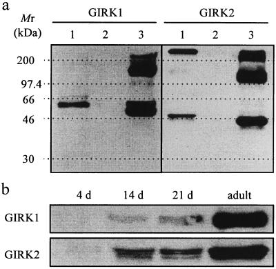
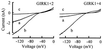
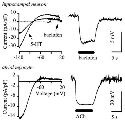
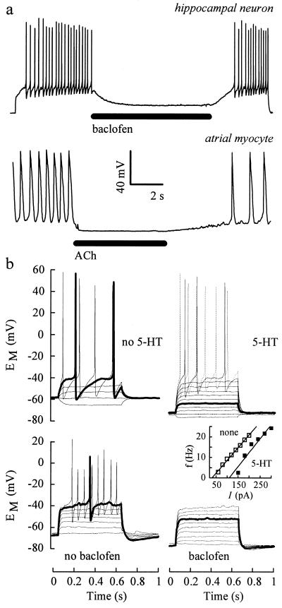
G protein-gated inward rectifier K+ channel subunits 1-4 (GIRK1-4) have been cloned from neuronal and atrial tissue and function as heterotetramers. To examine the inhibition of neuronal excitation by GIRKs, we overexpressed GIRKs in cultured hippocampal neurons from 18 day rat embryos, which normally lack or show low amounts of GIRK protein and currents. Adenoviral recombinants containing the cDNAs for GIRK1, GIRK2, GIRK4, and the serotonin 1A receptor were constructed. Typical GIRK currents could be activated by endogenous GABAB, serotonin 5-HT1A, and adenosine A1 receptors in neurons coinfected with GIRK1+2 or GIRK1+4. Under current clamp, GIRK activation increased the cell membrane conductance by 1- to 2-fold, hyperpolarized the cell by 11-14 mV, and inhibited action potential firing by increasing the threshold current for firing by 2- to 3-fold. These effects were not found in non- and mock-infected neurons, and were similar to the effects of muscarinic stimulation of native GIRK currents in atrial myocytes. Two inhibitory effects of GIRK activation, hyperpolarization and diminution of depolarizing pulses, were simulated from the experimental data. These inhibitory effects are physiologically important in the voltage range between the resting membrane potential and the potential where voltage-gated Na+ and K+ currents are activated; that is where GIRK currents are outward.


Figures

Figure 5
Figure 5
Simulations of the effects of gGIRK on cell membrane properties. (Upper Left) A simplified equivalent circuit incorporating only GIRK and leakage conductances. The membrane capacitance (Cm) is 50 pF. The leakage conductance (gleak, 55 pS/pF) is linear with a reversal potential of −12 mV (Eleak); gGIRK is as described in the text. (Upper Right) Current–voltage relation for the simulated gGIRK. The resting potential (−60 mV in the absence of GIRK activation) is simulated by an applied current of −132 pA in both panels. (Lower) Voltage changes in response to current injections (starting at 20 pA, 20 pA steps) with (Right) and without gGIRK (Left).
Figure 1
Figure 1
Expression of GIRK proteins in cultured rat E18 hippocampal neurons. (a) Overexpression of GIRK1 and GIRK2 proteins in cells coinfected with AdGIRK1+2. Total cellular protein was loaded (50 μg per lane): adult hippocampus (lane 1); 4 d-cultured neurons (lane 2); 4 d-cultured AdGIRK1+2-coinfected neurons, 3 d postinfection (lane 3). Left and right blots were probed with affinity-purified anti-GIRK1 and -GIRK2 antibodies, respectively. Protein samples were separated by SDS/PAGE with prestained molecular weight markers (Amersham) in neighboring lanes. The molecular weights of the marker proteins are indicated on the left with dotted lines across the gels. (b) Time-dependent expression of GIRK1 (Upper) and GIRK2 protein (Lower) in vitro. Total cellular protein of E18 neurons cultured for 4, 14, and 21 d, and of adult hippocampus was used (100 μg per lane) and probed with affinity-purified anti-GIRK1 and -GIRK2 antibodies, respectively.
Figure 2
Figure 2
Adenovirus-mediated expression of GIRK currents in hippocampal neurons. Cells cultured for 5 d were coinfected with AdGIRK1+2 or AdGIRK1+4 and analyzed 2 d postinfection in 25 mM [K+]o and 250 nM tetrodotoxin. Currents in the presence (b) and absence (a) of 100 μM baclofen; 0.5 mM Ba2+ blocked basal and baclofen-activated GIRK currents (c).
Figure 3
Figure 3
Cellular responses to GIRK activation at physiological [K+]o. (Left) Currents activated by 30 μM 5-HT and 50 μM baclofen in AdGIRK1+2-coinfected (4 d postinfection, thick lines) and uninfected (fine line, 5-HT only) hippocampal neurons 11 d in culture (Upper), and by 5 μM ACh in an atrial cell (Lower). (Right) EM changes caused by application of 50 μM baclofen and 5 μM ACh (bars); resting EM were −68 (neuron) and −24 mV (myocyte). Similar responses were observed to 8-OH-DPAT in neurons. The mean resting EM of 15 atrial cells was −35.8 ± 3.8 mV.
Figure 4
Figure 4
GIRK activation inhibits action potential firing. (a) Bars indicate the application of 50 μM baclofen to a 13 d-cultured neuron (3 d after coinfection with AdGIRK1+2), and of 5 μM ACh to an atrial cell. A 15.8 sec current pulse of +60 pA was injected into the neuron to cause firing; the myocyte was firing spontaneously. Resting EM were −70 mV (neuron) and −66 mV (myocyte). (b) EM responses of AdGIRK1+2-coinfected neurons to 0.6 sec current pulses in the presence (Right) and absence (Left) of 30 μM 5-HT (Upper) and 50 μM baclofen (Lower); steps of 20 pA (Upper, from −20 pA) and 16 pA (Lower, from 0 pA). Note that the 60 and 96 pA pulses cause action potential firing in the absence but not in the presence of 5-HT and baclofen, respectively (thick lines). (Inset) Firing frequencies calculated from action potential intervals of the top cell in the presence (▪) and absence (□) of 5-HT. Solid lines show linear fits; x axis intercepts indicate ITh.

Similar articles

Cited by

References

    1. Doupnik C A, Davidson N, Lester H A. Curr Opin Neurobiol. 1995;5:268–277. - PubMed
    1. Hedin K E, Lim N F, Clapham D E. Neuron. 1996;16:423–429. - PubMed
    1. Hille B. Ionic Channels of Excitable Membranes. Sunderland, MA: Sinauer; 1992.
    1. Loewi O. Pflügers Arch Gesamte Physiol. 1921;189:239–242.
    1. North R A. Br J Pharmacol. 1989;98:13–28. - PMC - PubMed

Publication types

MeSH terms

Substances

LinkOut - more resources