. 1997 Jun 30;137(7):1483-93.
doi: 10.1083/jcb.137.7.1483.

The lumenal domain of Sec63p stimulates the ATPase activity of BiP and mediates BiP recruitment to the translocon in Saccharomyces cerevisiae

Affiliations

The lumenal domain of Sec63p stimulates the ATPase activity of BiP and mediates BiP recruitment to the translocon in Saccharomyces cerevisiae

A K Corsi et al. J Cell Biol. .

Abstract

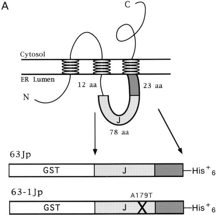
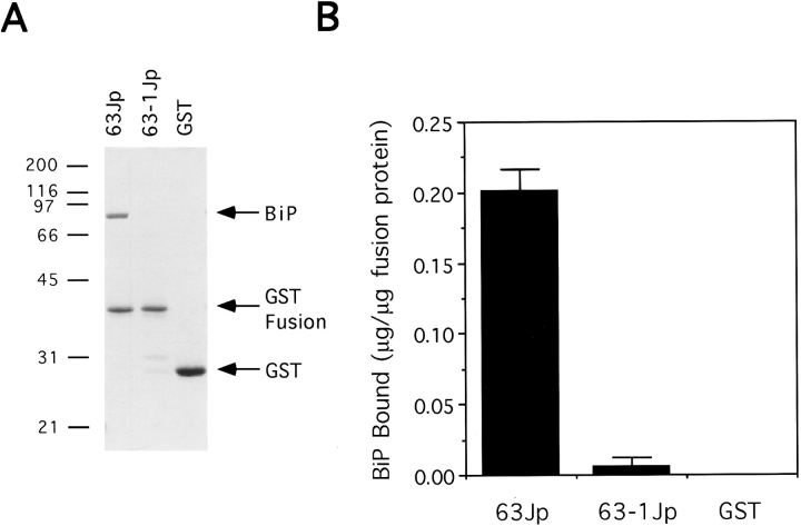
We studied the molecular nature of the interaction between the integral membrane protein Sec63p and the lumenal Hsp70 BiP to elucidate their role in the process of precursor transit into the ER of Saccharomyces cerevisiae. A lumenal stretch of Sec63p with homology to the Escherichia coli protein DnaJ is the likely region of interface between Sec63p and BiP. This domain, purified as a fusion protein (63Jp) with glutathione S-transferase (GST), mediated a stable ATP-dependent binding interaction between 63Jp and BiP and stimulated the ATPase activity of BiP. The interaction was highly selective because only BiP was retained on immobilized 63Jp when detergent-solubilized microsomes were mixed with ATP and the fusion protein. GST alone was inactive in these assays. Additionally, a GST fusion containing a point mutation in the lumenal domain of Sec63p did not interact with BiP. Finally, we found that the soluble Sec63p lumenal domain inhibited efficient precursor import into proteoliposomes reconstituted so as to incorporate both BiP and the fusion protein. We conclude that the lumenal domain of Sec63p is sufficient to mediate enzymatic interaction with BiP and that this interaction positioned at the translocation apparatus or translocon at the lumenal face of the ER is vital for protein translocation into the ER.


Figures

Figure 1
Figure 1
Construction and purification of the GST–Sec63p lumenal domain fusion protein. (A) The topology of Sec63p in the ER membrane is shown schematically according to Feldheim et al. (1992). The bottom portion of A depicts GST fused to either a wild-type Sec63p lumenal region (63Jp) or a mutant Sec63-1p lumenal region (63-1Jp) containing the A179T mutation found in sec63-1 (Nelson et al., 1993). (B) Fusions were purified as described in Materials and Methods. The peak fractions from the glutathione agarose and Ni2+-NTA agarose columns are shown on 12.5% SDS–polyacrylamide gels stained with Coomassie brilliant blue R-250.
Figure 1
Figure 1
Construction and purification of the GST–Sec63p lumenal domain fusion protein. (A) The topology of Sec63p in the ER membrane is shown schematically according to Feldheim et al. (1992). The bottom portion of A depicts GST fused to either a wild-type Sec63p lumenal region (63Jp) or a mutant Sec63-1p lumenal region (63-1Jp) containing the A179T mutation found in sec63-1 (Nelson et al., 1993). (B) Fusions were purified as described in Materials and Methods. The peak fractions from the glutathione agarose and Ni2+-NTA agarose columns are shown on 12.5% SDS–polyacrylamide gels stained with Coomassie brilliant blue R-250.
Figure 2
Figure 2
The Sec63p lumenal domain stimulates the ATPase of BiP. BiP (0.7 μg) was incubated at 25°C, pH 6.8, with increasing amounts of the Sec63p lumenal domain (63Jp) or the Sec63-1p lumenal domain (63-1Jp) fused to GST, or with GST alone. ATP hydrolysis (%) was measured by quantifying the percentage of 32Pi released from [γ-32P]ATP. All points reported represent 20 min of incubation at 25°C, which was determined to be in the linear range of the reactions.
Figure 3
Figure 3
BiP binds to the lumenal domain of Sec63p. (A) Glutathione agarose beads were first incubated with the proteins indicated at the top of the lanes and second with purified BiP in 1 mM ATP at pH 6.8. Beads were then washed as described in Materials and Methods, and the final eluate was resolved on a 12.5% SDS–polyacrylamide gel that was stained with Coomassie brilliant blue R-250. Molecular mass markers (in kD) are indicated at left. (B) BiP was quantified by comparing the signal shown with known amounts (not shown) using a scanner (model ScanMakerIII; Microtek, Redondo Beach, CA) and Imagequant v1.1 software (Molecular Dynamics).
Figure 4
Figure 4
The association of BiP and the Sec63p lumenal domain depends on the presence of hydrolyzable ATP. Binding reactions using 63Jp glutathione agarose beads were performed as in Fig. 3. (A) Final concentration of nucleotide in the reaction is indicated. The entire binding reaction depicted in lane 8 was done in the absence of Mg2+ plus 10 mM EDTA. (B) Bound nucleotides associated with glutathione beads (bound to either 63Jp or GST as indicated above the lanes) in the presence (+) or absence (−) of BiP after washing the beads were visualized by thin layer chromatography and a Phosphorimager.
Figure 4
Figure 4
The association of BiP and the Sec63p lumenal domain depends on the presence of hydrolyzable ATP. Binding reactions using 63Jp glutathione agarose beads were performed as in Fig. 3. (A) Final concentration of nucleotide in the reaction is indicated. The entire binding reaction depicted in lane 8 was done in the absence of Mg2+ plus 10 mM EDTA. (B) Bound nucleotides associated with glutathione beads (bound to either 63Jp or GST as indicated above the lanes) in the presence (+) or absence (−) of BiP after washing the beads were visualized by thin layer chromatography and a Phosphorimager.
Figure 5
Figure 5
BiP solubilized from yeast microsomes binds to 63Jp in an ATP- dependent manner. Soluble proteins from microsomes solubilized with Triton X-100 were mixed with a glutathione agarose 63Jp affinity matrix either with 1 mM ATP (+ ATP) or no ATP (− ATP) added. Binding reactions were at pH 6.8. The top panel is a 12.5% SDS–polyacrylamide gel stained with Coomassie brilliant blue R-250. Molecular mass markers (in kD) are on the left. P, insoluble microsomal proteins (pellet); FT, proteins not bound to affinity matrix (flow through); W, wash fractions; B, proteins still bound to beads after washing. The bottom panel is the region between 97 and 66 kD of an immunoblot probed with anti-BiP antibody.
Figure 6
Figure 6
63Jp binds selectively to BiP from detergent-solubilized microsomal membranes, and 63-1Jp associates with BiP from yeast microsomes in a pH-dependent manner. (A) 63Jp or 63-1Jp bound to glutathione agarose beads was incubated with solubilized proteins from a Triton X-100 detergent extract of wild-type yeast membranes in the presence of 1 mM ATP. The entire binding reaction was carried out at pH 6.8 or 8.0, as indicated. Protein profiles are shown on a 12.5% SDS–polyacrylamide gel stained with Coomassie brilliant blue R-250. All fractions are shown for 63Jp binding at pH 6.8; for the other conditions, only the eluate from the beads is shown. Molecular mass markers (in kD) are indicated on the left. L, microsomal detergent extract added to glutathione agarose beads; FT, protein not bound to beads after incubation with detergent lysate; W, material washed off the beads using binding buffer; B, proteins bound to glutathione agarose beads after washes. (B) To quantify the amount of BiP binding from the microsomal extracts, eluate fractions of binding assays were blotted to nitrocellulose and probed with anti-BiP antibody followed by 125I–protein A secondary antibody. The amount of BiP present in the eluate was then compared to a dilution series of the Triton X-100 extracts used in the experiment (not shown) to determine the percentage of BiP bound.
Figure 6
Figure 6
63Jp binds selectively to BiP from detergent-solubilized microsomal membranes, and 63-1Jp associates with BiP from yeast microsomes in a pH-dependent manner. (A) 63Jp or 63-1Jp bound to glutathione agarose beads was incubated with solubilized proteins from a Triton X-100 detergent extract of wild-type yeast membranes in the presence of 1 mM ATP. The entire binding reaction was carried out at pH 6.8 or 8.0, as indicated. Protein profiles are shown on a 12.5% SDS–polyacrylamide gel stained with Coomassie brilliant blue R-250. All fractions are shown for 63Jp binding at pH 6.8; for the other conditions, only the eluate from the beads is shown. Molecular mass markers (in kD) are indicated on the left. L, microsomal detergent extract added to glutathione agarose beads; FT, protein not bound to beads after incubation with detergent lysate; W, material washed off the beads using binding buffer; B, proteins bound to glutathione agarose beads after washes. (B) To quantify the amount of BiP binding from the microsomal extracts, eluate fractions of binding assays were blotted to nitrocellulose and probed with anti-BiP antibody followed by 125I–protein A secondary antibody. The amount of BiP present in the eluate was then compared to a dilution series of the Triton X-100 extracts used in the experiment (not shown) to determine the percentage of BiP bound.
Figure 7
Figure 7
The soluble Sec63p lumenal domain inhibits the ability of BiP to support ER translocation. (A) Proteoliposomes were made from kar2-159 microsomes with or without BiP added to 8% of total protein (12.8 μg). As indicated, in addition to BiP some proteoliposomes contained 63Jp added to 0.75% of total protein (0.5×; 1.2 μg) or either 63Jp, 63-1Jp, or GST added to 1.5% of total protein (1×; 2.4 μg). Translocation was assayed using [35S]methionine-labeled ppαF as a substrate. Aliquots from the translocation reactions were untreated (lane 1), treated with trypsin (lane 2), or treated with trypsin plus Triton X-100 (lane 3) and resolved on a 10% SDS–polyacrylamide gel. Translocation efficiency was determined using a Phosphorimager to calculate (pαf [lane 2] − pαf [lane 3])/(pαf + ppαF [lane 1]). Quantification is reported in B.
Figure 7
Figure 7
The soluble Sec63p lumenal domain inhibits the ability of BiP to support ER translocation. (A) Proteoliposomes were made from kar2-159 microsomes with or without BiP added to 8% of total protein (12.8 μg). As indicated, in addition to BiP some proteoliposomes contained 63Jp added to 0.75% of total protein (0.5×; 1.2 μg) or either 63Jp, 63-1Jp, or GST added to 1.5% of total protein (1×; 2.4 μg). Translocation was assayed using [35S]methionine-labeled ppαF as a substrate. Aliquots from the translocation reactions were untreated (lane 1), treated with trypsin (lane 2), or treated with trypsin plus Triton X-100 (lane 3) and resolved on a 10% SDS–polyacrylamide gel. Translocation efficiency was determined using a Phosphorimager to calculate (pαf [lane 2] − pαf [lane 3])/(pαf + ppαF [lane 1]). Quantification is reported in B.
Figure 8
Figure 8
A model for the interaction between Sec63p and BiP in posttranslational translocation across the ER membrane. (1) Our experiments suggest that BiP is recruited to the translocation apparatus by the lumenal domain of Sec63p. (2) The lumenal domain of Sec63p stimulates ATP hydrolysis by BiP to promote stable binding of BiP to the translocon. BiP may continue to associate with Sec63p while binding to the unfolded precursor protein emerging from the Sec61p pore. (3) The precursor or an unidentified protein then would catalyze nucleotide exchange in the ATP binding site of BiP, allowing BiP to undergo another cycle of interaction with Sec63p. This model does not include other precursor interactions with ER chaperones that may execute final secretory protein folding.

References

    1. Benaroudj N, Triniolles F, Ladjimi MM. Effect of nucleotides, peptides, and unfolded proteins on the self-association of the molecular chaperone HSC70. J Biol Chem. 1996;271:18471–18476. - PubMed
    1. Bernardi G. Chromatography of proteins on hydroxyapatite. Methods Enzymol. 1971;22:325–339. - PubMed
    1. Blond-Elguindi S, Fourie AM, Sambrook JF, Gething MH. Peptide-dependent stimulation of the ATPase activity of the molecular chaperone BiP is the result of conversion of oligomers to active monomers. J Biol Chem. 1993;268:12730–12735. - PubMed
    1. Brodsky JL. Post-translational protein translocation: not all hsc70s are created equal. Trends Biochem Sci. 1996;21:122–126. - PubMed
    1. Brodsky JL, Schekman R. A Sec63p-BiP complex from yeast is required for protein translocation in a reconstituted proteoliposome. J Cell Biol. 1993;123:1355–1363. - PMC - PubMed

Publication types

MeSH terms