. 1997 Jun 30;137(7):1511-24.
doi: 10.1083/jcb.137.7.1511.

The yeast v-SNARE Vti1p mediates two vesicle transport pathways through interactions with the t-SNAREs Sed5p and Pep12p

Affiliations

The yeast v-SNARE Vti1p mediates two vesicle transport pathways through interactions with the t-SNAREs Sed5p and Pep12p

G F von Mollard et al. J Cell Biol. .

Abstract

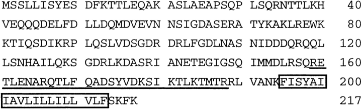
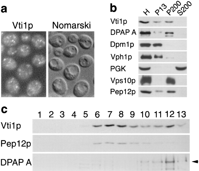
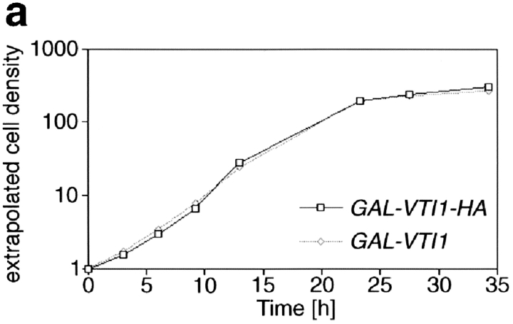
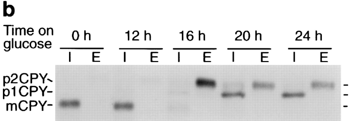
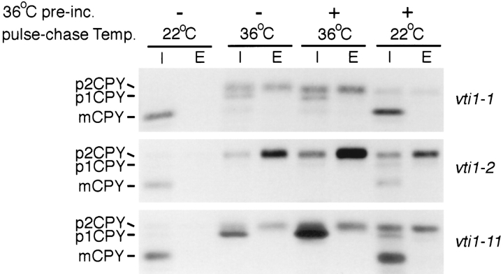
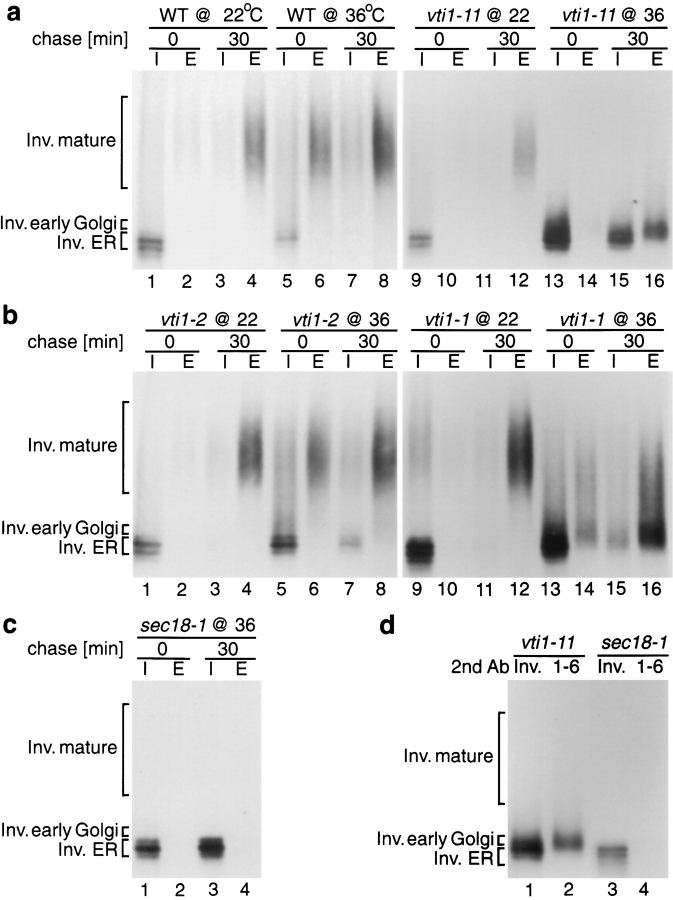
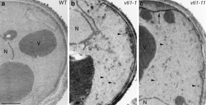
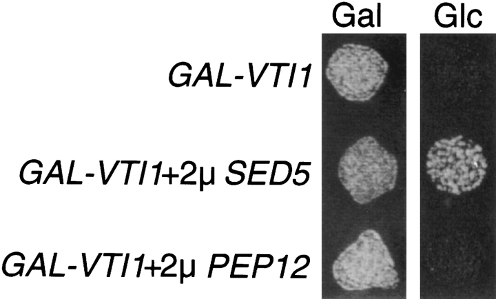
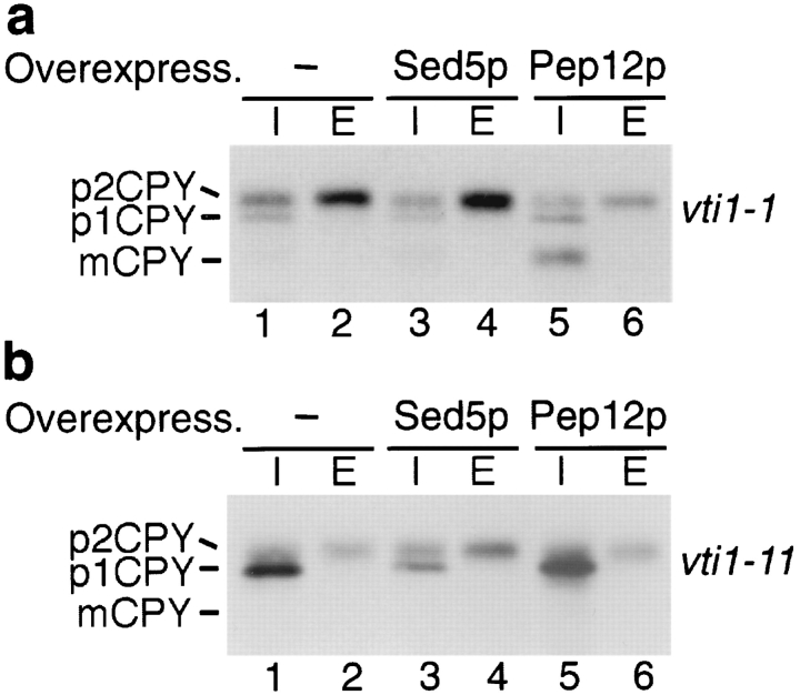
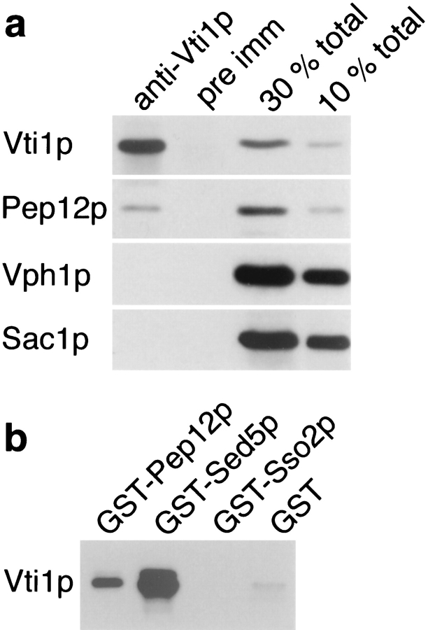
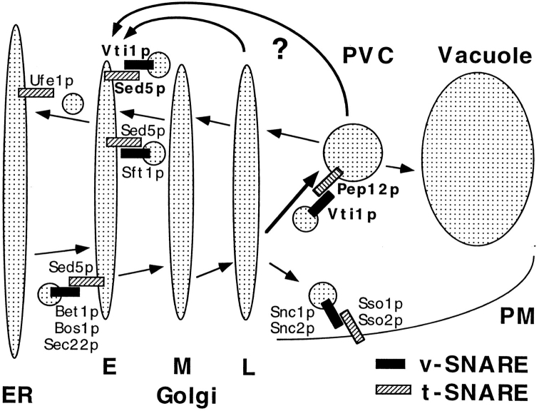
Membrane traffic in eukaryotic cells requires that specific v-SNAREs on transport vesicles interact with specific t-SNAREs on target membranes. We identified a novel Saccharomyces cerevisiae v-SNARE (Vti1p) encoded by the essential gene, VTI1. Vti1p interacts with the prevacuolar t-SNARE Pep12p to direct Golgi to prevacuolar traffic. vti1-1 mutant cells missorted and secreted the soluble vacuolar hydrolase carboxypeptidase Y (CPY) rapidly and reversibly when vti1-1 cells were shifted to the restrictive temperature. However, overexpression of Pep12p suppressed the CPY secretion defect exhibited by vti1-1 cells at 36 degrees C. Characterization of a second vti1 mutant, vti1-11, revealed that Vti1p also plays a role in membrane traffic at a cis-Golgi stage. vti1-11 mutant cells displayed a growth defect and accumulated the ER and early Golgi forms of both CPY and the secreted protein invertase at the nonpermissive temperature. Overexpression of the yeast cis-Golgi t-SNARE Sed5p suppressed the accumulation of the ER form of CPY but did not lead to CPY transport to the vacuole in vti1-11 cells. Overexpression of Sed5p allowed growth in the absence of Vti1p. In vitro binding and coimmunoprecipitation studies revealed that Vti1p interacts directly with the two t-SNAREs, Sed5p and Pep12p. These data suggest that Vti1p plays a role in cis-Golgi membrane traffic, which is essential for yeast viability, and a nonessential role in the fusion of Golgi-derived vesicles with the prevacuolar compartment. Therefore, a single v-SNARE can interact functionally with two different t-SNAREs in directing membrane traffic in yeast.


Figures

Figure 1
Figure 1
Amino acid sequence of yeast Vti1p. The predicted transmembrane domain is boxed. The predicted coiled coil domain is underlined (amino acids 159–188 with a P = 0.285 using the paircoil program [Berger et al., 1995]; amino acids 149–169 with a P = 0.58 using the coil program with a window of 21 [Lupas et al., 1991]). These sequence data are available from GenBank under accession number AF006074.
Figure 2
Figure 2
Vti1p localization. (a) Immunofluorescence microscopy of Vti1p. Anti-Vti1p antibodies reveal a punctate staining pattern distinct from the vacuoles seen by Nomarski optics. (b) Differential centrifugation of total yeast homogenate (H). Vti1p was predominantly found in the 200,000 g pellet (P200) with lower amounts in the 13,000 g pellet (P13). The syntaxin-related protein Pep12p had a similar distribution. (c) Sucrose density centrifugation of the S13 fraction. Vti1p migrated in two peaks, one cofractionating with the prevacuolar protein Pep12p and the other with the Golgi protein DPAP A. Fractions were collected from the top of the gradient (fraction 1) and analyzed by SDS-PAGE and immunoblotting.
Figure 3
Figure 3
Depletion of Vti1p leads to growth arrest and CPY mislocalization. (a) Growth curves of the GAL1–VTI1 and GAL1– VTI1-HA strains in glucose-containing medium. Cells of both strains displayed equivalent growth properties and stopped dividing after 22 h. Cell density was measured by optical density at 600 nm. The cultures were diluted when they reached an optical density of about one to keep cells growing in logarithmic phase. Taking the dilution factor into account, the extrapolated cell densities were calculated from the measured optical densities. (b) CPY sorting in the GAL1–VTI1-HA strain grown in galactose and for different amounts of time in glucose medium. CPY sorting was followed by pulse–chase labeling and CPY immunoprecipitation from cellular extracts (I, intracellular) and medium (E, extracellular). For cells cultured in galactose, CPY reached the vacuole (presence of mCPY). After 16 h in glucose most of the CPY was secreted as the late Golgi precursor form, p2CPY. After longer time periods in glucose, the ER and early Golgi forms of CPY (p1CPY) accumulated within the cell.
Figure 3
Figure 3
Depletion of Vti1p leads to growth arrest and CPY mislocalization. (a) Growth curves of the GAL1–VTI1 and GAL1– VTI1-HA strains in glucose-containing medium. Cells of both strains displayed equivalent growth properties and stopped dividing after 22 h. Cell density was measured by optical density at 600 nm. The cultures were diluted when they reached an optical density of about one to keep cells growing in logarithmic phase. Taking the dilution factor into account, the extrapolated cell densities were calculated from the measured optical densities. (b) CPY sorting in the GAL1–VTI1-HA strain grown in galactose and for different amounts of time in glucose medium. CPY sorting was followed by pulse–chase labeling and CPY immunoprecipitation from cellular extracts (I, intracellular) and medium (E, extracellular). For cells cultured in galactose, CPY reached the vacuole (presence of mCPY). After 16 h in glucose most of the CPY was secreted as the late Golgi precursor form, p2CPY. After longer time periods in glucose, the ER and early Golgi forms of CPY (p1CPY) accumulated within the cell.
Figure 4
Figure 4
CPY sorting in vti1-ts mutants. CPY sorting was followed in the vti1-1, vti1-2, and vti1-11 strains by pulse–chase labeling and CPY immunoprecipitations from cellular extracts (I, intracellular) and medium (E, extracellular). vti1-1 cells were preincubated for 30 min at 36°C and vti1-2 and vti1-11 cells for 15 min. In the vti1-1 and vti1-2 cells most CPY was secreted or accumulated intracellularly as p2CPY at the restrictive temperature. In vti1-11 cells, predominantly the ER or early Golgi form p1CPY accumulated intracellularly at the restrictive temperature.
Figure 5
Figure 5
Invertase sorting in vti1-ts mutants. (a–c) Invertase immunoprecipitations from intracellular (I) and extracellular (E) fractions were performed directly after the pulse or after a 30-min chase. Wild-type (WT), vti1, and sec18-1 cells were labeled at 22°C or after a 15-min preincubation at 36°C. Invertase was processed and secreted correctly in vti1-2 strains at either temperature. In vti1-1 cells, partially glycosylated invertase was secreted at 36°C. In vti1-11 cells, the ER and early Golgi forms of invertase accumulated, and partially glycosylated invertase was secreted at 36°C. (d) Invertase was immunoprecipitated from the intracellular fraction of vti1-11 and sec18-1 cells, which were incubated at 36°C and chased for 30 min. Secondary immunoprecipitations were performed with antiserum against invertase (Inv.) or against α1, 6–linked mannose (1-6). vti1-11 cells accumulated the ER form of invertase, which had not received α1,6–linked mannose, and the slightly larger cis-Golgi form, which could be precipitated with antiserum against α1,6–linked mannose.
Figure 6
Figure 6
Electron microscopy of wild-type and vti1 mutant cells. Cells were grown at 22°C and transferred to 37°C for 1 h. In vti1-1 cells (b), small vesicles (arrowheads) accumulated. In vti1-11 cells (c), small vesicles and ER membranes (arrow) accumulated. N, nucleus; V, vacuole. Bar, 500 nm.
Figure 7
Figure 7
SED5 overexpression suppresses vti1Δ lethality. Growth of the GAL1–VTI1 strain alone or overexpressing Sed5p or Pep12p on plates containing galactose (Gal) or glucose (Glc). The GAL1–VTI1 strain could not grow on glucose after the expression of VTI1 was shut off. Overexpression of the early Golgi t-SNARE Sed5p allowed growth in the absence of Vti1p. Overexpression of the prevacuolar-localized t-SNARE Pep12p did not support growth of vti1Δ cells.
Figure 8
Figure 8
CPY sorting in vti1-1 (a) and vti1-11 (b) cells overexpressing Sed5p and Pep12p. Overexpression of Pep12p in vti1-1 cells partially restored CPY sorting to the vacuole at the restrictive temperature (36°C). Overexpression of Sed5p in vti1-11 mutant cells reduced p1CPY accumulation at the restrictive temperature.
Figure 9
Figure 9
Physical interaction of Vti1p with Sed5p and Pep12p. (a) Coimmunoprecipitation of Pep12p with Vti1p. Yeast spheroplasts were solubilized with 0.5% Triton X-100. Vti1p was quantitatively precipitated from the Triton extract by native immunoprecipitation with anti-Vti1p antibodies, and ∼10% of the total Pep12p was coimmunoprecipitated. In contrast, the membrane proteins Vph1p (vacuole) and Sac1p (ER and Golgi apparatus) were not coimmunoprecipitated, and pre-immune serum did not precipitate any of these proteins. For quantitative comparison, Triton extracts equivalent to 30 and 10% of total protein present in the immunoprecipitations were analyzed in lanes 3 and 4. The proteins bound to the beads and the starting extracts were analyzed by SDS-PAGE and immunoblotting with antibodies against Vti1p, Pep12p, Vph1p, and Sac1p. (b) In vitro binding of the soluble domain of Vti1p to immobilized GST fusion proteins containing the soluble domains of different yeast t-SNAREs. Vti1p interacted with GST-Sed5p and GST-Pep12p but not with GST-Sso2p or GST alone. Equal amounts of immobilized GST fusion proteins were incubated with 6-His–Vti1p. The bound proteins were analyzed by SDS-PAGE and immunoblotting with anti-Vti1p antibodies.
Figure 10
Figure 10
Model for Vti1p function in vesicle docking and fusion. Yeast Golgi- derived vesicles contain the v-SNARE Vti1p on the membrane allowing for its interaction with the t-SNARE Pep12p on the surface of the prevacuolar compartment (PVC). This interaction provides part of the specificity for fusion of Golgi-derived vesicles with the prevacuole. In addition, Vti1p interacts with the early Golgi t-SNARE Sed5p. We propose that Vti1p is involved in retrograde traffic from either the prevacuolar compartment or the late Golgi compartment back to the early Golgi compartment. Other v-SNAREs and t-SNAREs functioning in yeast ER, Golgi, and plasma membrane (PM) traffic are included. E, early Golgi; M, medial Golgi; L, late Golgi; PVC, prevacuolar compartment; PM, plasma membrane.

References

    1. Aalto MK, Ronne H, Keranen S. Yeast syntaxins Sso1p and Sso2p belong to a family of related membrane proteins that function in vesicular transport. EMBO (Eur Mol Biol Organ) J. 1993;12:4095–4104. - PMC - PubMed
    1. Banfield DK, Lewis MJ, Pelham HR. A SNARE-like protein required for traffic through the Golgi complex. Nature (Lond) 1995;375:806–809. - PubMed
    1. Bankaitis VA, Johnson LM, Emr SD. Isolation of yeast mutants defective in protein targeting to the vacuole. Proc Natl Acad Sci USA. 1986;83:9075–9079. - PMC - PubMed
    1. Bartel PL, Fields S. Analyzing protein-protein interactions using two-hybrid system. Methods Enzymol. 1995;254:241–263. - PubMed
    1. Becherer KA, Rieder SE, Emr SD, Jones EW. Novel syntaxin homolog, Pep12p, required for the sorting of lumenal hydrolases to the lysosome-like vacuole in yeast. Mol Biol Cell. 1996;7:579–594. - PMC - PubMed

Publication types

MeSH terms

Associated data