. 1997 Jun 30;137(7):1537-53.
doi: 10.1083/jcb.137.7.1537.

The Chlamydomonas mating type plus fertilization tubule, a prototypic cell fusion organelle: isolation, characterization, and in vitro adhesion to mating type minus gametes

Affiliations

The Chlamydomonas mating type plus fertilization tubule, a prototypic cell fusion organelle: isolation, characterization, and in vitro adhesion to mating type minus gametes

N F Wilson et al. J Cell Biol. .

Abstract

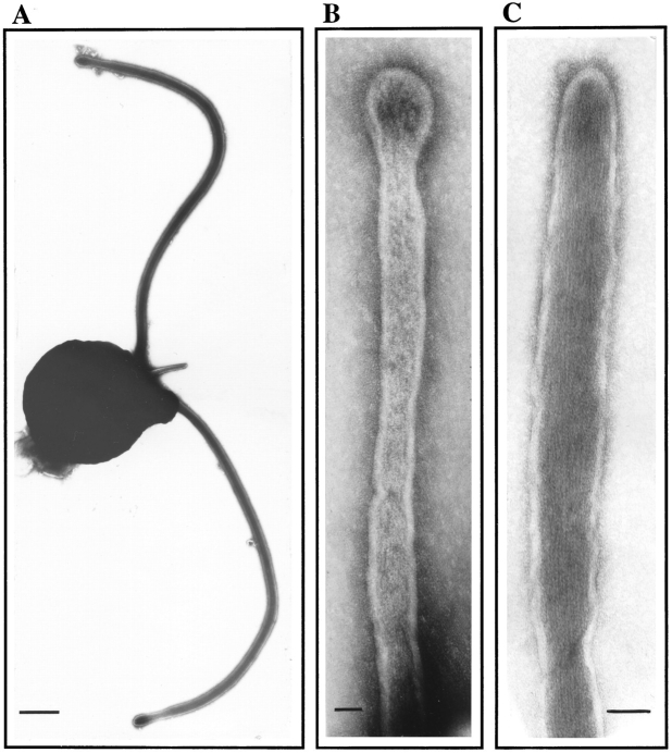
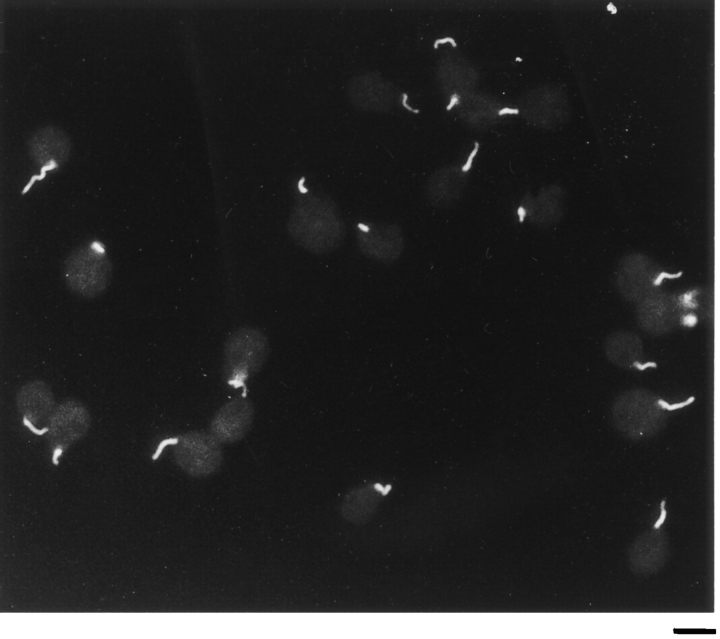
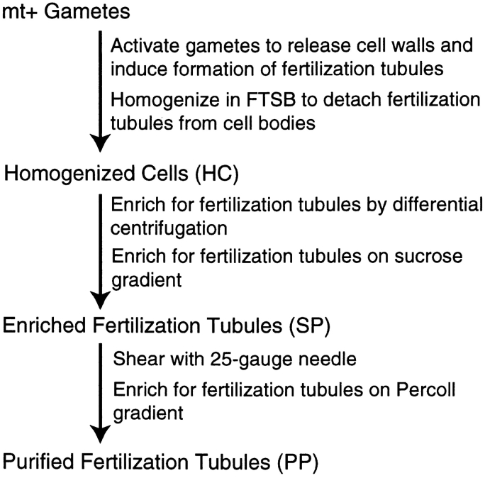
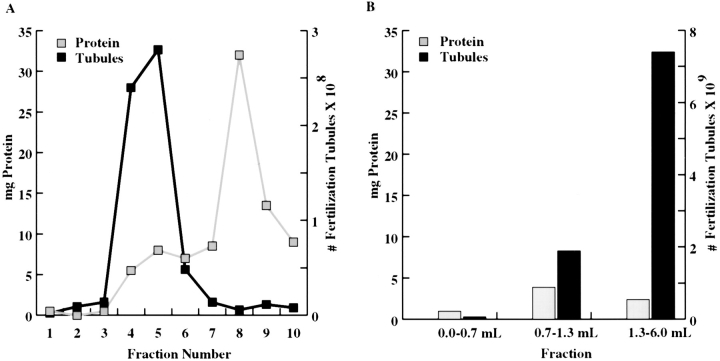
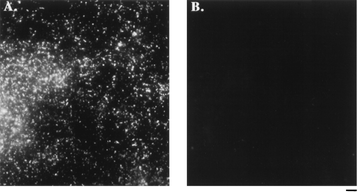
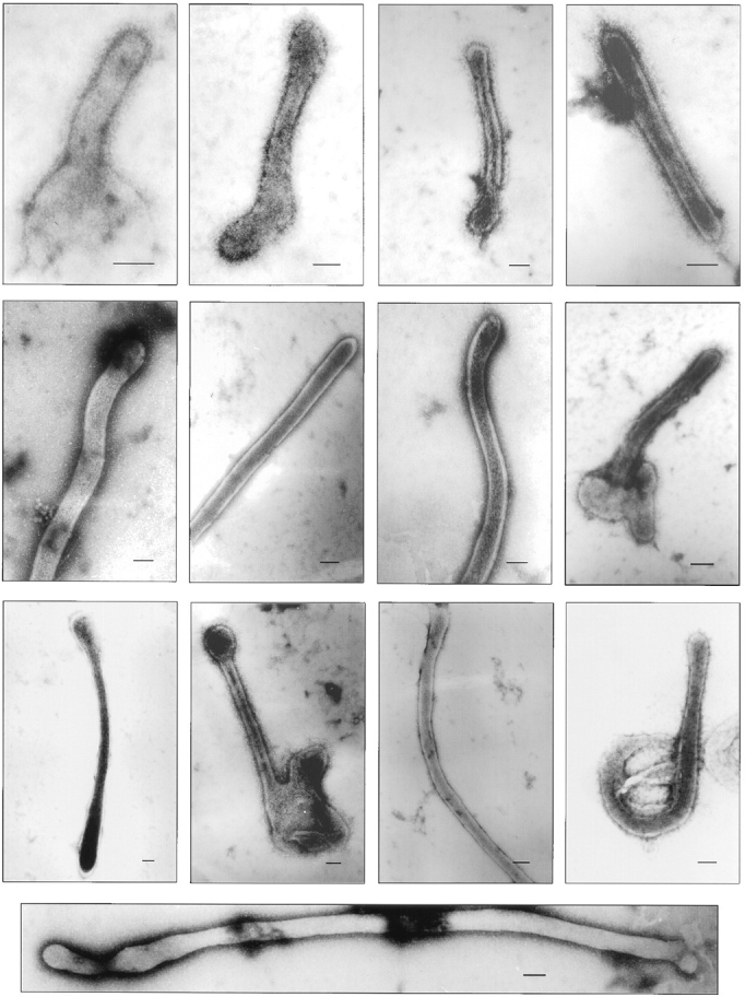
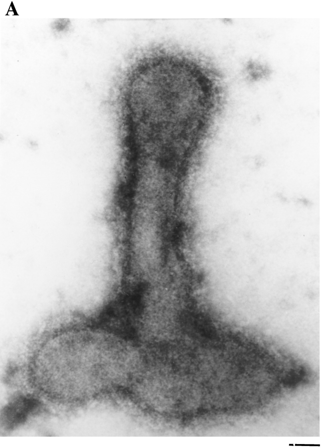
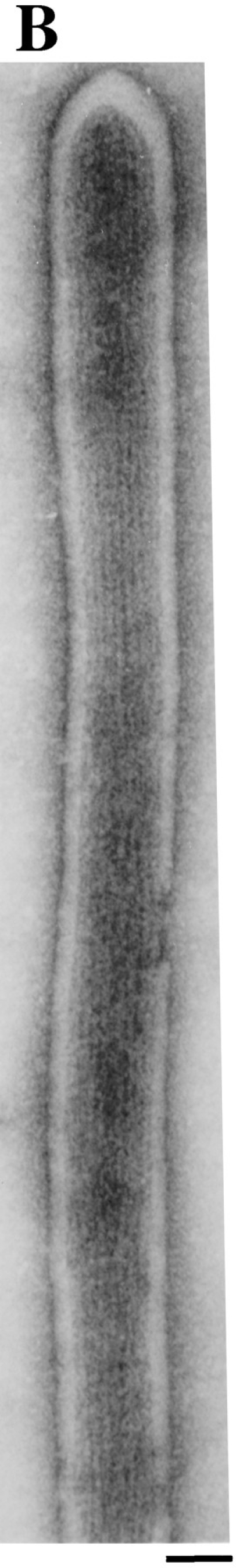
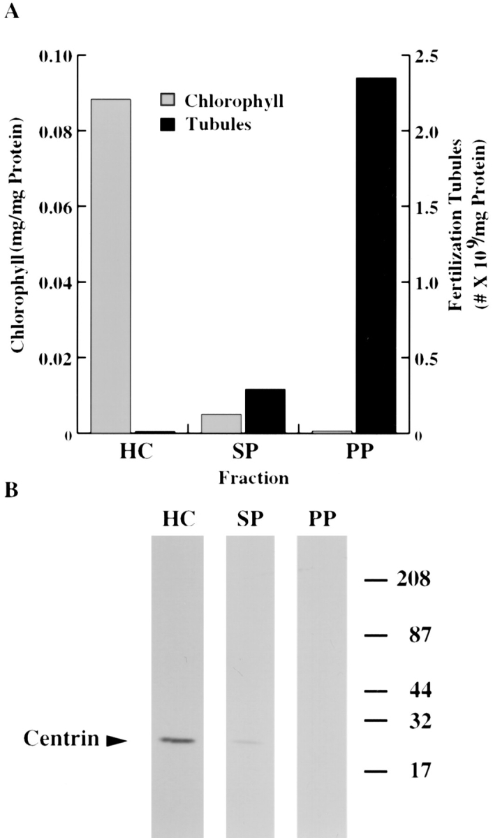
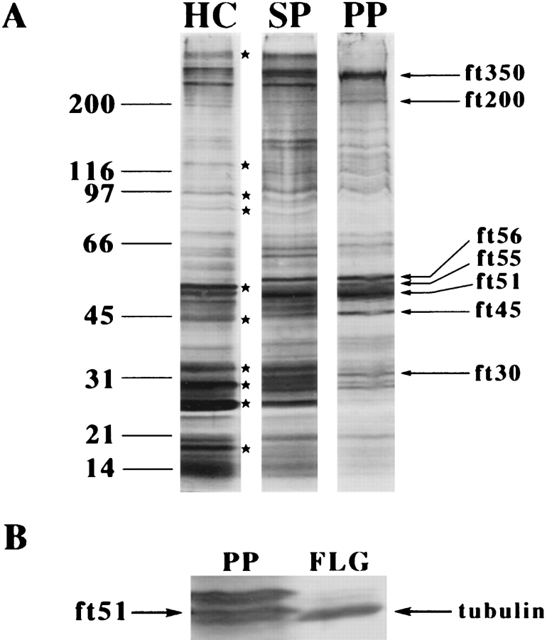
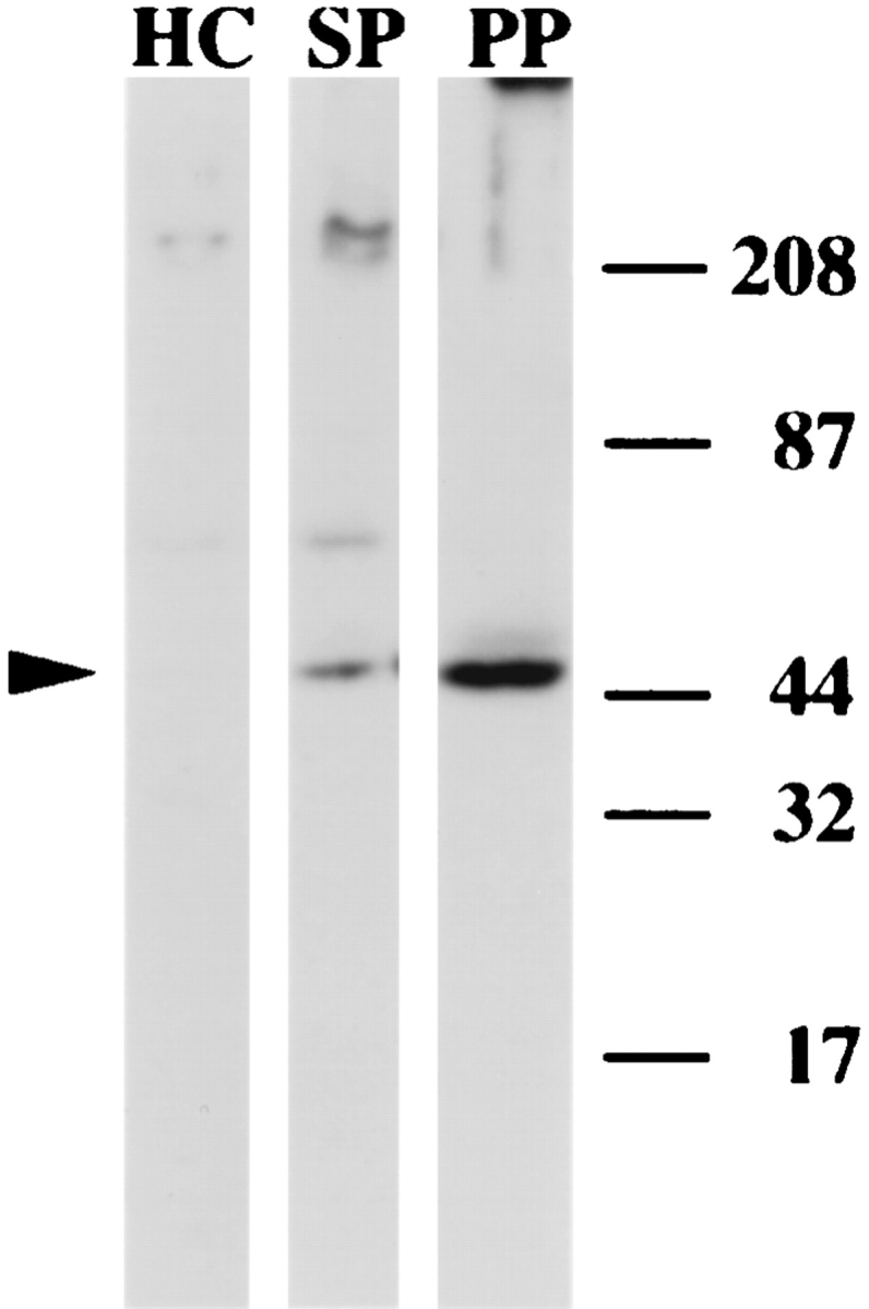
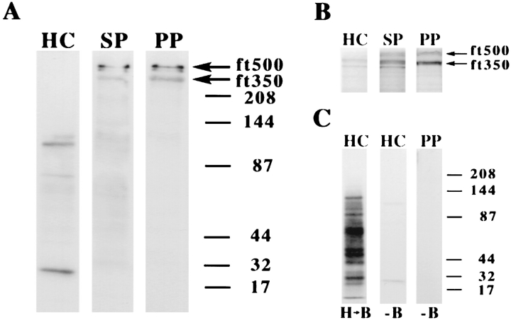
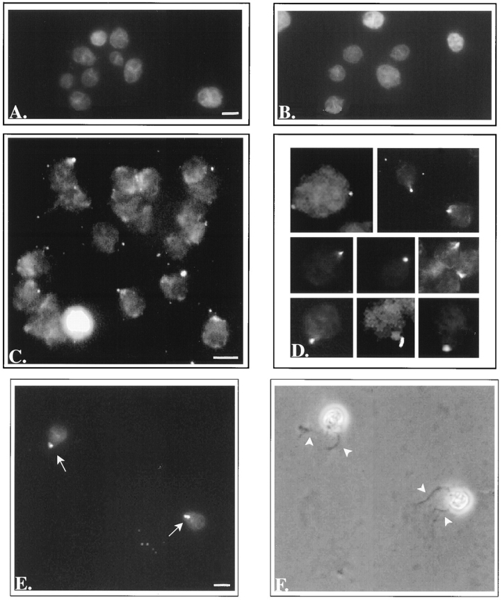
In the biflagellated alga Chlamydomonas, adhesion and fusion of the plasma membranes of gametes during fertilization occurs via an actin-filled, microvillus-like cell protrusion. Formation of this approximately 3-microm-long fusion organelle, the Chlamydomonas fertilization tubule, is induced in mating type plus (mt+) gametes during flagellar adhesion with mating type minus (mt-) gametes. Subsequent adhesion between the tip of the mt+ fertilization tubule and the apex of a mating structure on mt- gametes is followed rapidly by fusion of the plasma membranes and zygote formation. In this report, we describe the isolation and characterization of fertilization tubules from mt+ gametes activated for cell fusion. Fertilization tubules were detached by homogenization of activated mt+ gametes in an EGTA-containing buffer and purified by differential centrifugation followed by fractionation on sucrose and Percoll gradients. As determined by fluorescence microscopy of samples stained with a fluorescent probe for filamentous actin, the method yielded 2-3 x 10(6) fertilization tubules/microg protein, representing up to a 360-fold enrichment of these organelles. Examination by negative stain electron microscopy demonstrated that the purified fertilization tubules were morphologically indistinguishable from fertilization tubules on intact, activated mt+ gametes, retaining both the extracellular fringe and the internal array of actin filaments. Several proteins, including actin as well as two surface proteins identified by biotinylation studies, copurified with the fertilization tubules. Most importantly, the isolated mt+ fertilization tubules bound to the apical ends of activated mt- gametes between the two flagella, the site of the mt- mating structure; a single fertilization tubule bound per cell, binding was specific for gametes, and fertilization tubules isolated from trypsin-treated, activated mt+ gametes did not bind to activated mt- gametes.


Figures

Figure 1
Figure 1
Negative stain electron microscopy of fertilization tubules on activated mt+ gametes. mt+ gametes activated by mixing with an equal number of mt− gametes were negatively stained and examined by electron microscopy. (A) Low magnification view of an activated mt+ gamete with a fertilization tubule extending from the apical region of the cell between the two flagella. (B and C) Higher magnification views of fertilization tubules on intact mt+ gametes. Bars: (A) 1 μm; (B and C) 0.05 μm.
Figure 2
Figure 2
Bodipy phallacidin staining of activated mt+ gametes. mt+ gametes were activated with dibutyryl cAMP and papaverine, and fertilization tubules were visualized by fluorescent microscopy after staining with bodipy phallacidin. Bar, 5 μm.
Figure 3
Figure 3
Diagrammatic representation of the method for isolation of fertilization tubules.
Figure 4
Figure 4
Enrichment of fertilization tubules by fractionation on sucrose and Percoll gradients. (A) Crude preparations of fertilization tubules from differential centrifugation were fractionated on 15–60% sucrose gradients as described in Materials and Methods. After centrifugation, 0.5-ml fractions were collected from above, and the numbers of fertilization tubules/μg protein in each fraction were determined (gray line, protein; black line, fertilization tubules). (B) Fractions 4 and 5 from the sucrose gradients in A were pooled, collected by centrifugation, and further fractionated on 30% Percoll gradients as described in Materials and Methods. Fractions of the indicated volumes were collected from above, and the numbers of fertilization tubules/μg protein in each fraction were determined (gray bars, protein; black bars, fertilization tubules).
Figure 5
Figure 5
Bodipy phallacidin staining of purified fertilization tubules. Isolated fertilization tubules were harvested from the Percoll gradient fraction enriched in fertilization tubules, fixed, and diluted 1:80 with FTSB before staining with bodipy phallacidin. Large numbers of fertilization tubules were visible in the fluorescein channel (A), while no obvious chloroplast contamination was observed in the rhodamine channel (B). Bar, 5 μm.
Figure 6
Figure 6
Negative stain electron microscopic analysis at low magnification of isolated fertilization tubules. Isolated fertilization tubules were harvested from the Percoll gradient fraction enriched in fertilization tubules, fixed, stained as described in Materials and Methods, and examined by electron microscopy. Actin filaments and fringe are visible in these images of isolated fertilization tubules. Bars, 0.05 μm.
Figure 7
Figure 7
Negative stain electron microscopic analysis at high magnification of isolated fertilization tubules. (A) High magnification view of an isolated fertilization tubule showing the extracellular fringe on the distal portion of the fertilization tubule. (B) Enlargement of an isolated fertilization tubule from the panel in Fig. 6, showing the large number of actin filaments in this structure. Bars, 0.05 μm.
Figure 7
Figure 7
Negative stain electron microscopic analysis at high magnification of isolated fertilization tubules. (A) High magnification view of an isolated fertilization tubule showing the extracellular fringe on the distal portion of the fertilization tubule. (B) Enlargement of an isolated fertilization tubule from the panel in Fig. 6, showing the large number of actin filaments in this structure. Bars, 0.05 μm.
Figure 8
Figure 8
De-enrichment of chlorophyll and centrin. (A) The amounts of chlorophyll present at various steps in the isolation of fertilization tubules (homogenized cells [HC], sucrose pellets [SP], and Percoll pellets [PP]) were determined as described in Materials and Methods (gray bars, chlorophyll; black bars, fertilization tubules). (B) Immunoblot analysis with anticentrin monoclonal antibody, 17E10, of various steps in the isolation of fertilization tubules. Samples (12 μg) of homogenized cells (lane HC), sucrose pellets (lane SP), and Percoll pellets (lane PP) were separated by SDS-PAGE and analyzed by immunoblotting as described. The location of centrin is indicated by the arrowhead on the left. The migration of prestained molecular weight markers is shown on the right.
Figure 9
Figure 9
SDS-PAGE analysis of fertilization tubule proteins. (A) Samples (12 μg) of homogenized cells (lane HC), sucrose pellets (lane SP), and Percoll pellets (lane PP) were separated by SDS-PAGE and stained with silver. Asterisks indicate proteins that de-enriched, and arrows indicate proteins that enriched during isolation of fertilization tubules. The migration of unstained molecular weight markers is indicated on the left. (B) Migration of ft51 above flagellar tubulin on SDS-PAGE. Samples of purified fertilization tubules (12 μg, lane PP) and isolated mt+ gametic flagella (∼3 μg, lane FLG) were analyzed by SDS-PAGE and silver staining as described.
Figure 10
Figure 10
Enrichment of actin during purification of fertilization tubules. Samples (12 μg) of homogenized cells (lane HC), sucrose pellets (lane SP), and Percoll pellets (lane PP) were separated by SDS-PAGE and analyzed by immunoblotting with an anti-Volvox actin polyclonal antibody as described. The location of actin is indicated by the arrowhead on the left. Prestained molecular weight markers are shown on the right.
Figure 11
Figure 11
Identification of surface proteins by vectorial labeling with biotin. (A) Activated mt+ gametes were labeled with Sulfo-NHS-Biotin, fertilization tubules were isolated, and samples (12 μg) of homogenized cells (lane HC), sucrose pellets (lane SP), and Percoll pellets (lane PP) were separated by SDS-PAGE and analyzed by streptavidin blotting. Arrows indicate the locations of surface-biotinylated proteins of 500 (ft500) and 350 kD (ft350). (B) Staining with Coomassie blue of the same samples as in A. Arrows indicate the locations of ft500 and ft350. (C) To identify proteins biotinylated in disrupted cells, activated mt+ gametes were homogenized before biotinylation (lane HB) and 1.5 μg of protein was analyzed by SDS-PAGE and streptavidin blotting. In addition, potential streptavidin-binding proteins were identified by analyzing both nonbiotinylated homogenized cells (12 μg, HC, −B) and fertilization tubules purified from nonbiotinylated cells (12 μg, PP, −B).
Figure 12
Figure 12
Binding of fertilization tubules to activated mt− gametes. (A) Fluorescence microscopy of bodipy phallacidin stained mt− gametes activated and fixed as described in Materials and Methods and incubated with FTSB alone. (B) De-walled, fixed, mt− vegetative cells incubated with isolated fertilization tubules. (C and D) Activated, fixed mt− gametes incubated with isolated fertilization tubules. (E) Fluorescence and (F) phase contrast micrographs of activated, live mt− gametes incubated with isolated fertilization tubules. E and F are images of the same cells. Arrows point to the fertilization tubules visible in the fluorescent image shown in E, and arrowheads in F indicate the apically located flagella on the same cells. Bars (for pairs A and B, C and D, and E and F), 5 μm.

Similar articles

Cited by

References

    1. Adair WS, Monk BC, Cohen R, Hwang C, Goodenough UW. Sexual agglutinins from the Chlamydomonasflagellar membrane: partial purification and characterization. J Biol Chem. 1982;257:4593–4602. - PubMed
    1. Bergman K, Goodenough UW, Goodenough DA, Jawitz J, Martin H. Gametic differentiation in Chlamydomonas reinhardtii.II. Flagellar membranes and the agglutination reaction. J Cell Biol. 1975;67:606–622. - PMC - PubMed
    1. Berlin C, Bargatze RF, Campbell JJ, von Andrian UH, Szabo MC, Hasslen SR, Nelson RD, Berg EL, Erlandsen SL, Butcher EC. α4 integrins mediate lymphocyte attachment and rolling under physiologic flow. Cell. 1995;80:413–422. - PubMed
    1. Berryman M, Gary R, Bretscher A. Ezrin oligomers are major cytoskeletal components of placental microvilli: a proposal for their involvement in cortical morphogenesis. J Cell Biol. 1995;131:1231–1242. - PMC - PubMed
    1. Blobel CP, Wolfsberg TG, Turck CW, Myles DG, Primakoff P, White JM. A potential fusion peptide and an integrin ligand domain in a protein active in sperm-egg fusion. Nature (Lond) 1992;356:248–252. - PubMed

Publication types