. 1997 Jun 30;137(7):1555-66.
doi: 10.1083/jcb.137.7.1555.

IQGAP1, a Rac- and Cdc42-binding protein, directly binds and cross-links microfilaments

Affiliations

IQGAP1, a Rac- and Cdc42-binding protein, directly binds and cross-links microfilaments

A M Bashour et al. J Cell Biol. .

Abstract

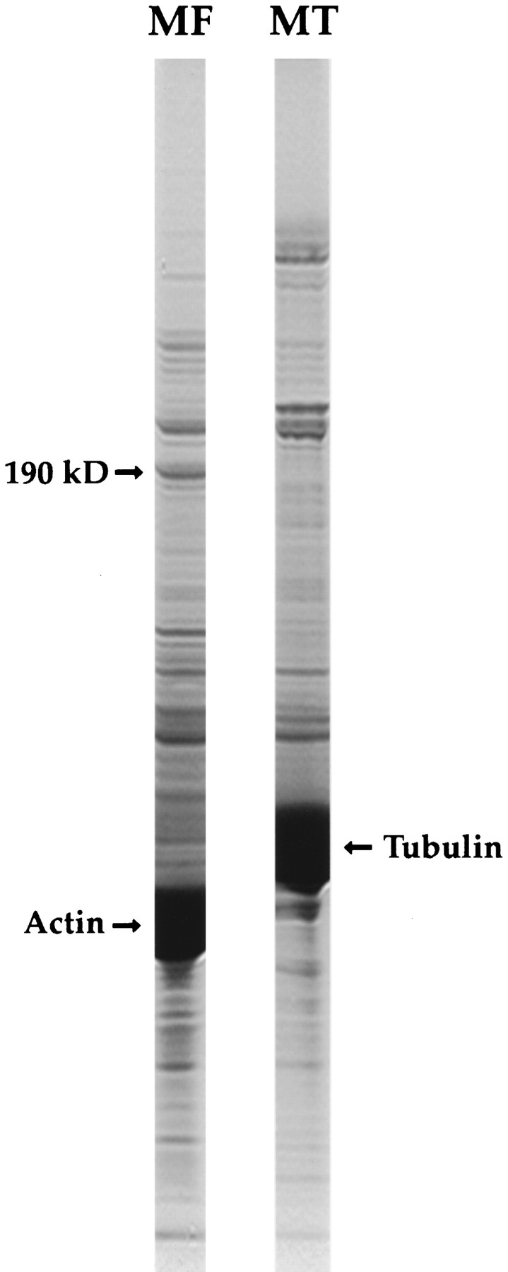
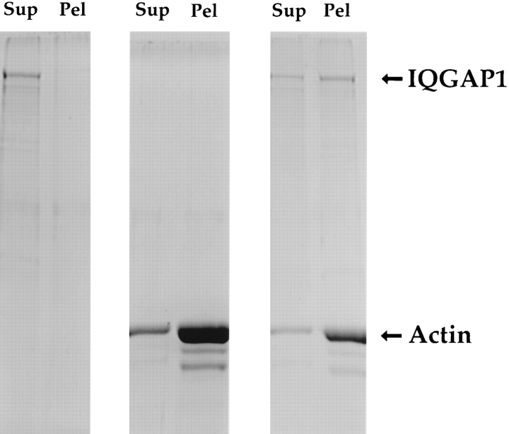
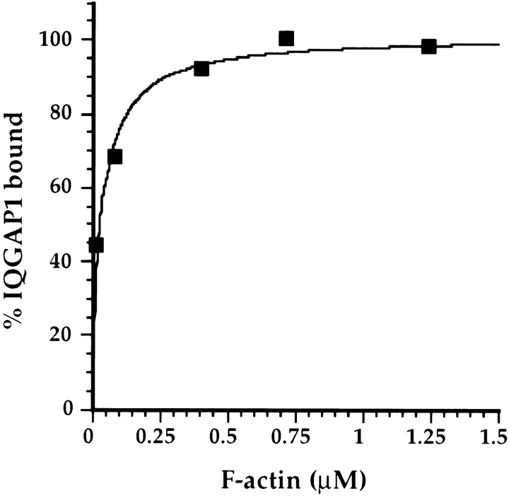
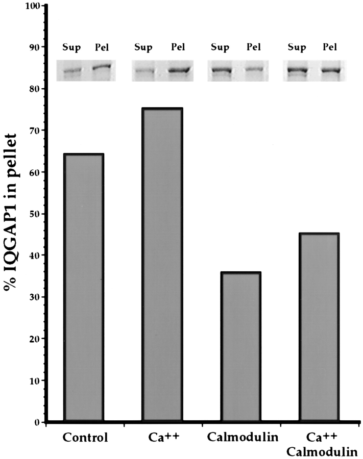
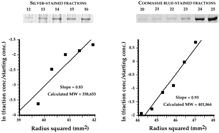
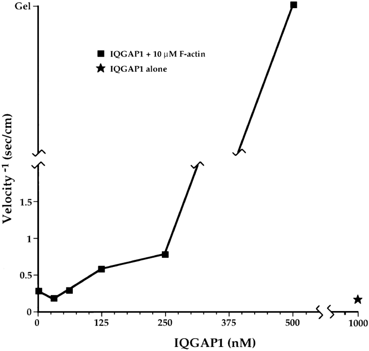
Activated forms of the GTPases, Rac and Cdc42, are known to stimulate formation of microfilament-rich lamellipodia and filopodia, respectively, but the underlying mechanisms have remained obscure. We now report the purification and characterization of a protein, IQGAP1, which is likely to mediate effects of these GTPases on microfilaments. Native IQGAP1 purified from bovine adrenal comprises two approximately 190-kD subunits per molecule plus substoichiometric calmodulin. Purified IQGAP1 bound directly to F-actin and cross-linked the actin filaments into irregular, interconnected bundles that exhibited gel-like properties. Exogenous calmodulin partially inhibited binding of IQGAP1 to F-actin, and was more effective in the absence, than in the presence of calcium. Immunofluorescence microscopy demonstrated cytochalasin D-sensitive colocalization of IQGAP1 with cortical microfilaments. These results, in conjunction with prior evidence that IQGAP1 binds directly to activated Rac and Cdc42, suggest that IQGAP1 serves as a direct molecular link between these GTPases and the actin cytoskeleton, and that the actin-binding activity of IQGAP1 is regulated by calmodulin.


Figures

Figure 1
Figure 1
A 190-kD putative actin-binding protein is present in bovine adrenal tissue. MFs assembled from purified rabbit muscle actin, or MTs assembled with taxol from purified bovine brain tubulin, were added to bovine adrenal cytosol at 4°C. After centrifugation, the MT and MF pellets were resuspended and analyzed by SDS-PAGE. As can be seen, a prominent 190-kD polypeptide was present exclusively in the MF pellet.
Figure 4
Figure 4
Purified IQGAP1 binds directly to F-actin. To account for the presence of IQGAP1 in MF pellets isolated from adrenal cytosol (see Fig. 1), IQGAP1 could have associated directly or indirectly with F-actin. To distinguish between these possibilities, purified IQGAP1 at 0.15 mg/ml was centrifuged alone (left) or in combination with 0.14 mg/ml (3.2 μM) F-actin (right), and the supernatants (Sup) and pellets (Pel) were analyzed by SDS-PAGE. As a control, F-actin alone at 0.28 mg/ml (6.4 μM) was also centrifuged in parallel (center). As shown here, purified IQGAP1 bound directly to F-actin.
Figure 5
Figure 5
IQGAP1 binds F-actin with high affinity. 0.15 mg/ml IQGAP1 was mixed with various amounts of F-actin, and the samples were centrifuged. The pellets were then resuspended to the initial sample volumes, and the amounts of IQGAP1 and F-actin in each pellet were determined by quantitative SDS-PAGE. A plot of the percentage of IQGAP1 bound vs F-actin is shown here, and the data were fitted to a hyperbolic curve. In this experiment, two-thirds of the total IQGAP1 was able to bind F-actin, and 0.1 μg/ml IQGAP1 was therefore defined as 100% bound. Note that 50% of the binding-competent IQGAP1 was bound at ∼40 nM F-actin. When the data were reanalyzed by the Scatchard method, a curved or multiphasic plot was obtained (not shown), implying the existence of multiple classes of binding sites on IQGAP1 for F-actin. The fact that half of the IQGAP1 bound to 40 nM F-actin should thus be regarded as an indication of high affinity binding, but 40 nM is probably not a reliable estimate of the dissociation constant.
Figure 6
Figure 6
Calmodulin inhibits binding of IQGAP1 to F-actin. 0.19 mg/ml IQGAP1 (1 μM 190K subunit; 4 μM IQ domains) plus 10 μM F-actin were centrifuged in the absence of additional factors, or in the presence of calcium (∼250 μM above the EGTA concentration), 40 μM calmodulin (160 μM calcium binding sites), or calcium plus calmodulin. Note that 64% of the IQGAP1 pelleted in the absence of other factors, while binding in the presence of calcium rose slightly to 75%. Binding was potently inhibited by calmodulin, and demonstrably, but less so, by calcium plus calmodulin. Only 36% of the total IQGAP1 bound to F-actin in the presence of calmodulin alone, while 45% bound in the presence of calmodulin plus calcium.
Figure 2
Figure 2
The 190-kD polypeptide is equivalent to IQGAP1. The 190-kD polypeptide was purified in denatured form by preparative SDS-PAGE, electroeluted from the gel, and digested in solution with endoproteinase lys C. Four peptides were purified by HPLC and sequenced. Comparison of the peptide sequences with the GenBank/EMBL/DDBJ data bank indicated high identity of the bovine adrenal protein with human IQGAP1, a 1,658–amino acid residue protein that contains four potential calmodulin-binding IQ domains, and a region homologous to catalytic domains of GAPs (40). Sequences corresponding to peptides 1 and 4 are also found in human IQGAP2, a protein which is 62% identical to IQGAP1, but is reportedly abundant only in liver (8). A 9-mer peptide corresponding to the highlighted residues within peptide 3 was used as an immunogen for the production of an anti-IQGAP1 antibody.
Figure 7
Figure 7
Two 190-kD polypeptides are present in each native IQGAP1 molecule. Sedimentation equilibrium in a table top ultracentrifuge was used to determine the molecular weight of native purified IQGAP1 (see Materials and Methods for details). Quantitative densitometry of a silver-stained gel of the five least dense fractions (left) and of a Coomassie blue– stained gel of the six densest fractions (right) yielded data that were plotted as indicated on the two graphs. The slope of each graph is a function of molecular weight, which was calculated to be 358,653 and 401,864 for the silver and Coomassie stains, respectively. The average of these two figures is 380,256, and the data are most readily explained by a native IQGAP1 molecule that comprises two ∼190-kD subunits.
Figure 3
Figure 3
Purified, native IQGAP1 contains copurifying, but substoichiometic calmodulin. (A) Native IQGAP1 was purified from bovine adrenal cytosol by FPLC using sequential cation exchange (S-Sepharose), gel filtration (Sephacryl S-300), and anion exchange (Q-Sepharose) columns. A Coomassie blue–stained gel of 5-μg samples of cytosol and each of the three column steps are shown in the upper part of the figure. The lower part of the figure illustrates corresponding Western blots for the 190-kD IQGAP1 subunit and calmodulin, as well as a calmodulin concentration series (8, 16, and 32 ng). Note that the 190-kD subunit was undetectable in cytosol at the exposure shown here, and that it became progressively enriched throughout the purification procedure. Note also that 5 μg of purified IQGAP1 contained ∼20 ng of calmodulin, or an ∼1:20 molar ratio of calmodulin to the 190-kD polypeptide. (B) To determine whether the calmodulin present in purified IQGAP1 represented a copurifying subunit of the native molecule or a trace contaminant, Q-Sepharose fractions that contained the 190-kD polypeptide were analyzed by Western blotting with anticalmodulin. Coelution of the 190-kD polypeptide and calmodulin were observed, although the calmodulin peak was more skewed than the 190-kD peak toward late-eluting fractions (see Discussion). Calmodulin thus appears to be a bona fide, albeit substoichiometric subunit of purified native IQGAP1.
Figure 10
Figure 10
IQGAP1 colocalizes with cytochalasin D–sensitive microfilaments in lamellipodia and ruffles, but not in stress fibers. CV-1 cells were stained with a rabbit antibody to IQGAP1 (17) followed by rhodamine-labeled goat anti–rabbit IgG and Bodipy–phallicidin (to visualize F-actin). Intense IQGAP1 and F-actin staining was colocalized in lamellipodia located at cell margins (top row), and in ruffles located on the upper surfaces of some cells (middle row). Weak, diffuse staining by anti-IQGAP1 was also seen throughout the cytoplasm, and may represent soluble protein. Stress fibers, which contain densely packed MFs and were brightly stained by Bodipy–phallicidin, were not stained by anti-IQGAP1. After a 30-min exposure to 5 μg/ml cytochalasin D, which caused F-actin to depolymerize, cells were substantially depleted of IQGAP1-positive structures, and of stress fibers as well (bottom row). Bar, 10 μm.
Figure 8
Figure 8
Purified IQGAP1 causes F-actin to gel. F-actin at 10 μM was mixed with various concentrations of purified IQGAP1 ranging from 0–500 nM, and each sample was analyzed by falling ball viscometry (see Materials and Methods for details). Note that the ball was able to travel though a mixture of 10 μM actin plus 250 nM IQGAP1, but that a gel-like state, in which the ball did not move, was achieved at 500 nM IQGAP1. Note also that the ball moved readily through a sample of 1 μM IQGAP1 in the absence of F-actin.
Figure 9
Figure 9
Purified IQGAP1 cross-links F-actin into bundles. Negative stain EM was used to analyze F-actin alone (1.6 μM polymerized actin) or in combination with purified IQGAP1 (0.4 μM). Striking bundles of F-actin were observed when IQGAP1 was present. The bundles were of variable diameter and length, and often appeared to form intricate interconnected branches. IQGAP1 alone did not form bundles (not shown). Bar, 200 nm.

Similar articles

Cited by

References

    1. Amano M, Chihara K, Kimura K, Fukata Y, Nakamura N, Matsuura Y, Kaibuchi K. Formation of actin stress fibers and focal adhesions enhanced by Rho-kinase. Science (Wash DC) 1997;275:1308–1311. - PubMed
    1. Andre E, Lottspeich F, Schleicher M, Noegel A. Severin, gelsolin, and villin share a homologous sequence in regions presumed to contain F-actin severing domains. J Biol Chem. 1988;263:722–727. - PubMed
    1. Bagrodia S, Derijard B, Davis RJ, Cerione RA. Cdc42 and PAK-mediated signaling leads to Jun kinase and p38 mitogen-activated protein kinase activation. J Biol Chem. 1995;270:27995–27998. - PubMed
    1. Blanchard A, Ohanian V, Critchley D. The structure and function of alpha-actinin. J Muscle Res Cell Motil. 1989;10:280–289. - PubMed
    1. Bloom GS, Wagner MC, Pfister KK, Brady ST. Native structure and physical properties of bovine brain kinesin and identification of the ATP-binding subunit polypeptide. Biochemistry. 1988;27:3409–3416. - PubMed

Publication types

Substances