. 1997 Jun 30;137(7):1639-49.
doi: 10.1083/jcb.137.7.1639.

ZAP-70 protein tyrosine kinase is constitutively targeted to the T cell cortex independently of its SH2 domains

Affiliations

ZAP-70 protein tyrosine kinase is constitutively targeted to the T cell cortex independently of its SH2 domains

R D Huby et al. J Cell Biol. .

Abstract

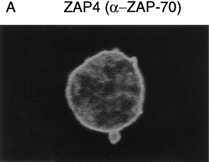
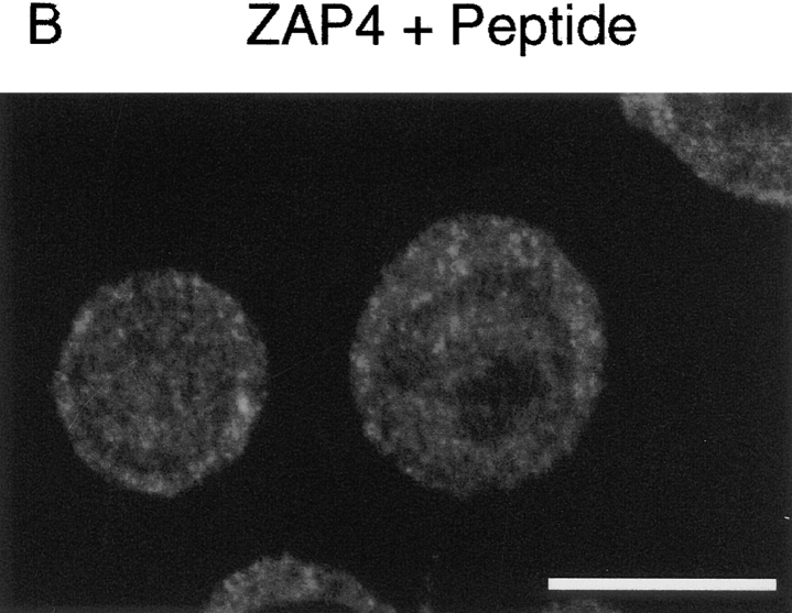
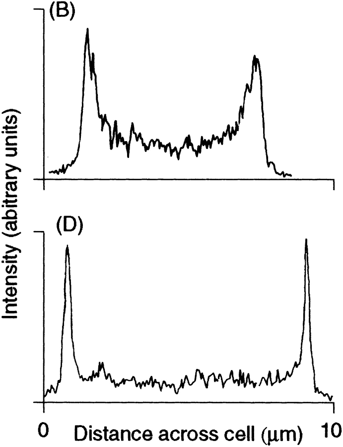
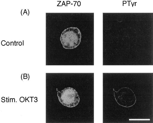
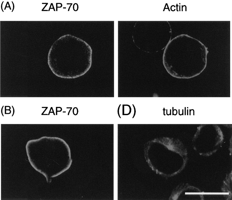
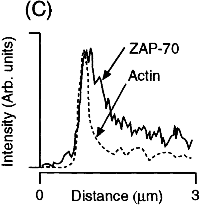
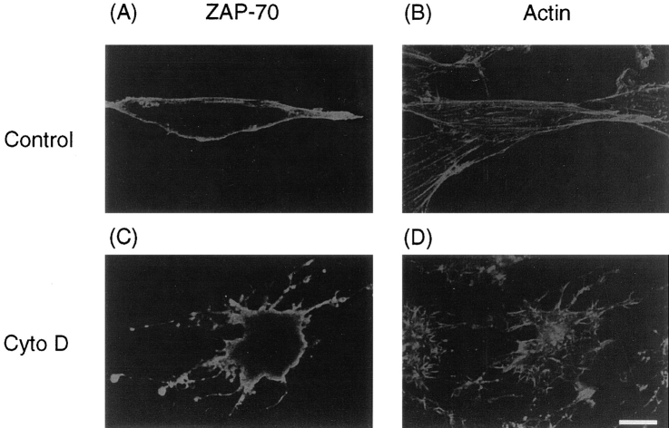
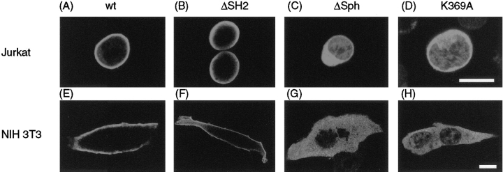
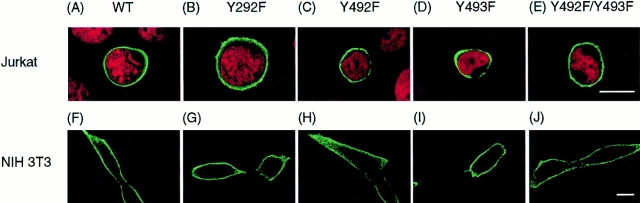
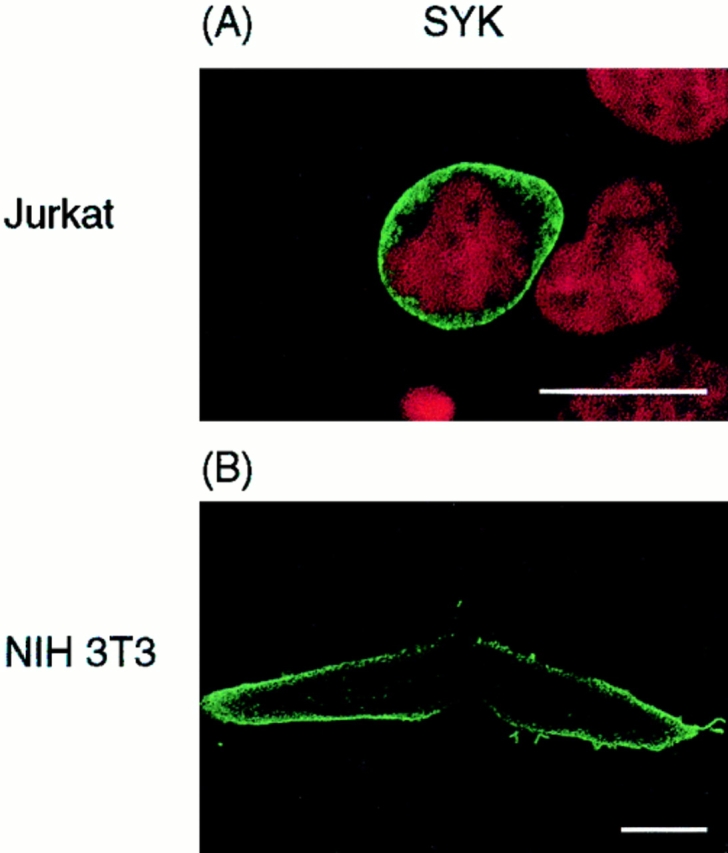
ZAP-70 is a nonreceptor protein tyrosine kinase that is essential for signaling via the T cell antigen receptor (TCR). ZAP-70 becomes phosphorylated and activated by LCK protein tyrosine kinase after interaction of its two NH2-terminal SH2 domains with tyrosine-phosphorylated subunits of the activated TCR. In this study, the localization of ZAP-70 was investigated by immunofluorescence and confocal microscopy. ZAP-70 was found to be localized to the cell cortex in a diffuse band under the plasma membrane in unstimulated T cells, and this localization was not detectably altered by TCR stimulation. Analysis of mutants indicated that ZAP-70 targeting was independent of its SH2 domains but required its active kinase domain. The specific compartmentalization of ZAP-70 suggests that it may interact with an anchoring protein in the cell cortex via its hinge or kinase domains. It is likely that the maintenance of high concentrations of ZAP-70 at the cell cortex, that only has to move a short distance to interact with phophorylated TCR subunits, facilitates rapid initiation of signaling by the TCR. In addition, as the major increase in tyrosine phosphorylation induced by the TCR also occurs at the cell cortex (Ley, S.C., M. Marsh, C.R. Bebbington, K. Proudfoot, and P. Jordan. 1994. J. Cell. Biol. 125:639-649), ZAP-70 may be localized close to its downstream targets.


Figures

Figure 1
Figure 1
Localization of endogenous ZAP-70 in Jurkat T cells. Staining with ZAP 4 anti–ZAP-70 antiserum in Jurkat T cells reveals specific cortical staining (A) which was abrogated by the addition of blocking peptide, to which the antiserum was raised during the primary staining step (B). Low levels of nonspecific staining were also detected in the cytoplasm. Bar, 10 μm.
Figure 1
Figure 1
Localization of endogenous ZAP-70 in Jurkat T cells. Staining with ZAP 4 anti–ZAP-70 antiserum in Jurkat T cells reveals specific cortical staining (A) which was abrogated by the addition of blocking peptide, to which the antiserum was raised during the primary staining step (B). Low levels of nonspecific staining were also detected in the cytoplasm. Bar, 10 μm.
Figure 2
Figure 2
Localization of HA-tagged ZAP-70 after transient transfection into Jurkat T cells. Transfected cells were double stained with anti-HA tag mAb 12CA5 (green) and the nuclear stain 7–amino-actinomycin D (red), revealing that ZAP-70–HA expression is limited primarily to the cell cortex, with maximal staining proximal to the plasma membrane (A). A gradient of ZAP-70–HA staining intensity measured according to pixel brightness was quantified along a cell transect as shown in (B). In comparison, very bright LCK staining is seen after transient transfection with an LCK expression plasmid, which nevertheless remains tightly and symmetrically associated with the plasma membrane (C), illustrated quantitatively by the corresponding cell transect (D). Transfected ZAP-70–HA is shown to behave similarly to endogenous ZAP-70 after TCR activation, becoming tyrosine phosphorylated and associated with phosphorylated TCR ζ (E). Bar, 10 μm.
Figure 2
Figure 2
Localization of HA-tagged ZAP-70 after transient transfection into Jurkat T cells. Transfected cells were double stained with anti-HA tag mAb 12CA5 (green) and the nuclear stain 7–amino-actinomycin D (red), revealing that ZAP-70–HA expression is limited primarily to the cell cortex, with maximal staining proximal to the plasma membrane (A). A gradient of ZAP-70–HA staining intensity measured according to pixel brightness was quantified along a cell transect as shown in (B). In comparison, very bright LCK staining is seen after transient transfection with an LCK expression plasmid, which nevertheless remains tightly and symmetrically associated with the plasma membrane (C), illustrated quantitatively by the corresponding cell transect (D). Transfected ZAP-70–HA is shown to behave similarly to endogenous ZAP-70 after TCR activation, becoming tyrosine phosphorylated and associated with phosphorylated TCR ζ (E). Bar, 10 μm.
Figure 3
Figure 3
ZAP-70 is largely soluble before fixation. Jurkat T cells were disrupted by Dounce homogenization (A) and separated into three fractions; (P1) pellet recovered by 280 g centrifugation, comprised primarily of nuclear debris and the actin cytoskeleton; (P2) pellet recovered at 100,000 g comprised membranous fragments, protein aggregates, and the remaining soluble fraction S1. ZAP-70 compartmentalization was unaffected by stimulation of cells for 5 or 20 min with OKT3 F(ab)2 before homogenization. After fixation for 5 min in 2% paraformaldehyde, essentially all ZAP-70 became insoluble (B) and was recovered in the pellet P rather than the soluble fraction S after centrifugation for 2 min at 8,000 g.
Figure 4
Figure 4
Localization of ZAP-70 is unaffected by TCR stimulation. Jurkat T cells were either unstimulated (A), or stimulated for 5 min with F(ab)2 fragments of the CD3 mAb, OKT3 (B), fixed, and double stained for endogenous ZAP-70 (left) and total phosphotyrosine (right). Staining for endogenous ZAP-70 with ZAP-4 antiserum also produced cytoplasmic fluorescence which was nonspecific (Fig. 1). Bar, 10 μm.
Figure 5
Figure 5
ZAP-70 does not colocalize with actin or α-tubulin. ZAP-70–HA transfected Jurkat T cells were double stained to reveal ZAP–70 and actin microfilaments in the same cell (A). ZAP-70– HA gave a broader staining pattern, quantified by the pixel density transect shown in B. Double staining was similarly used to reveal ZAP-70–HA and α-tubulin distribution within the same cells (C). Bar, 10 μm.
Figure 5
Figure 5
ZAP-70 does not colocalize with actin or α-tubulin. ZAP-70–HA transfected Jurkat T cells were double stained to reveal ZAP–70 and actin microfilaments in the same cell (A). ZAP-70– HA gave a broader staining pattern, quantified by the pixel density transect shown in B. Double staining was similarly used to reveal ZAP-70–HA and α-tubulin distribution within the same cells (C). Bar, 10 μm.
Figure 6
Figure 6
ZAP-70 localization is unaffected by disruption of the actin cytoskeleton. NIH 3T3 fibroblasts were transfected with a plasmid encoding ZAP-70–HA, fixed, and double stained to reveal the distribution of ZAP-70–HA (A) and actin microfilaments (B) in the same cells. Treatment of cells with cytochalasin D before fixation did not delocalize ZAP-70–HA from the cell cortex (C), although the actin cytoskeleton was disrupted (D). Bar, 10 μm.
Figure 7
Figure 7
ZAP-70 requires an active kinase domain for its localization. Jurkat T cells (A–D) or NIH 3T3 fibroblasts (E–H) were transfected with plasmids encoding either: wild-type ZAP-70–HA (A and E), ΔSH2-ZAP-70–HA (B and F), ΔSph-ZAP-70–HA (C and G), or kinase-dead ZAP-70.K369A–HA. Transfected cells were stained with 12CA5 to reveal the HA-tagged ZAP-70 protein. Bars, 10 μm.
Figure 8
Figure 8
Inducibly phosphorylated tyrosine residues are not involved in ZAP-70 localization to the cortex. Jurkat T cells (A–E) or NIH 3T3 fibroblasts (F–J) were transfected with plasmids encoding either wild-type ZAP-70 (A and F), Y292F (B and G), Y492F (C and H), Y493F (D and I), or Y492F/Y493F (E and H). Unlike previous experiments, all constructs were HA-tagged at the NH2 terminus. Transfected cells were stained with 12CA5 mAb to reveal HA-tagged ZAP-70 protein (green) and in the case of Jurkat were counterstained with 7–amino-actinomycin D to reveal the position of the nucleus (red). Bars, 10 μm.
Figure 9
Figure 9
SYK localizes to the cell cortex. Jurkat T cells (A) or 3T3 fibroblasts (B) were transfected with a plasmid encoding NH2 terminally HA-tagged SYK, fixed, and the localization of the expressed protein determined, as described in Fig. 7. Bars, 10 μm.

Similar articles

Cited by

References

    1. Appleby MW, Gross JA, Cooke MP, Levin SD, Qian X, Perlmutter RM. Defective T cell receptor signaling in mice lacking the thymic isoform of p59fyn. Cell. 1992;70:751–763. - PubMed
    1. Baniyash M, Garcia-Morales P, Luong E, Samelson LE, Klausner RD. The T cell antigen receptor ζ chain is tyrosine phosphorylated upon activation. J Biol Chem. 1988;263:18225–18230. - PubMed
    1. Bu JY, Shaw AS, Chan AC. Analysis of the interaction of ZAP-70 and syk protein-tyrosine kinases with the T-cell antigen receptor by plasmon resonance. Proc Natl Acad Sci USA. 1995;92:5106–5110. - PMC - PubMed
    1. Chan AC, Shaw AS. Regulation of antigen receptor signal transduction by protein tyrosine kinases. Current Opin Immunol. 1996;8:394–401. - PubMed
    1. Chan AC, Iwashima M, Turck CW, Weiss A. ZAP-70: a 70 kd protein-tyrosine kinase that associates with the TCR zeta chain. Cell. 1992;71:649–662. - PubMed

Publication types

Substances