. 1997 Jun 30;137(7):1651-62.
doi: 10.1083/jcb.137.7.1651.

Dynamics of beta-catenin interactions with APC protein regulate epithelial tubulogenesis

Affiliations

Dynamics of beta-catenin interactions with APC protein regulate epithelial tubulogenesis

A L Pollack et al. J Cell Biol. .

Abstract

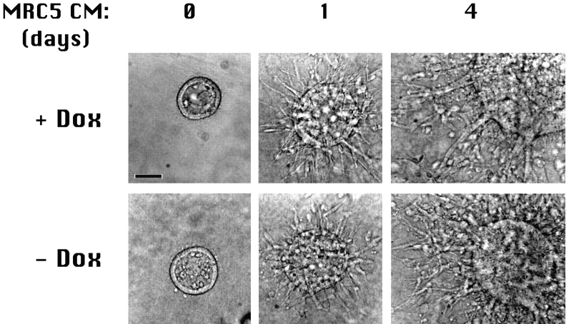
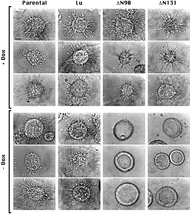
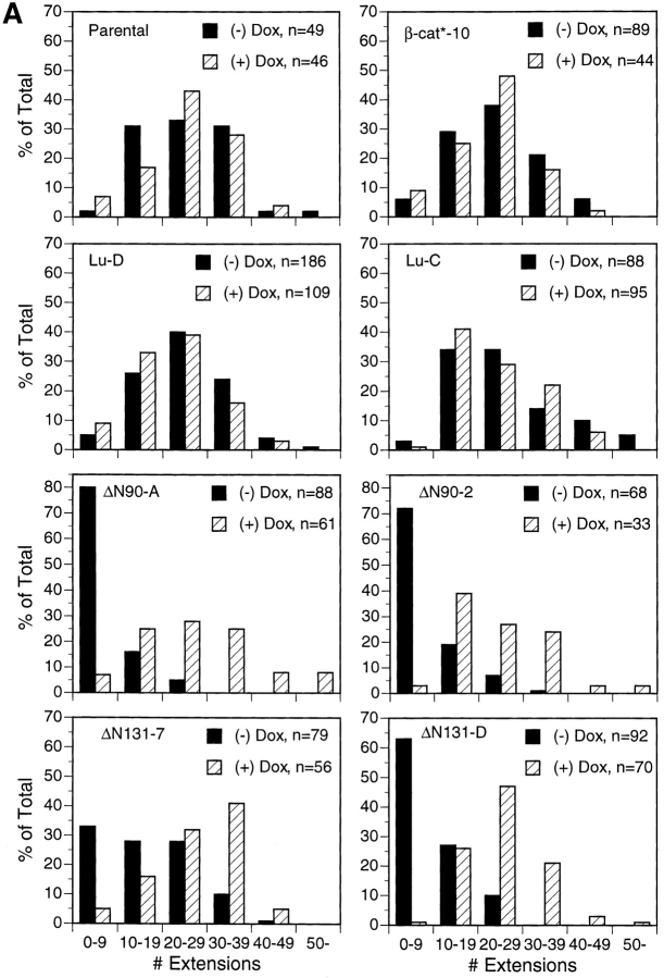
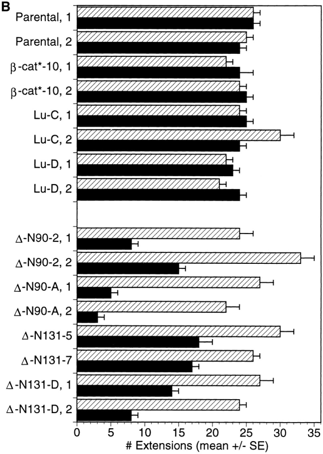
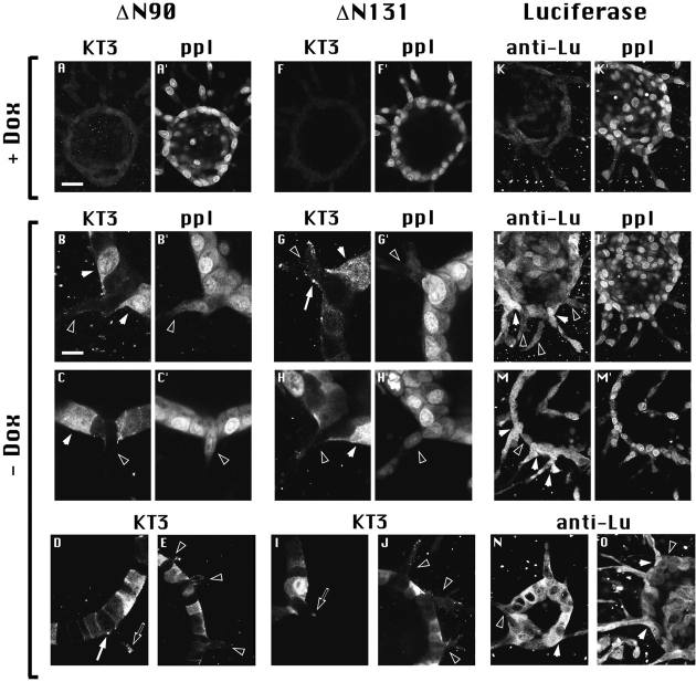
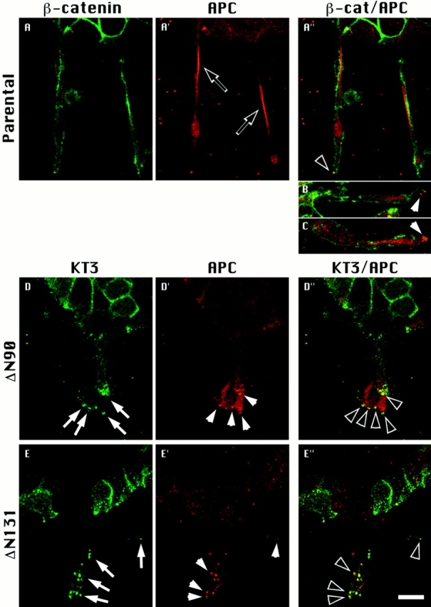
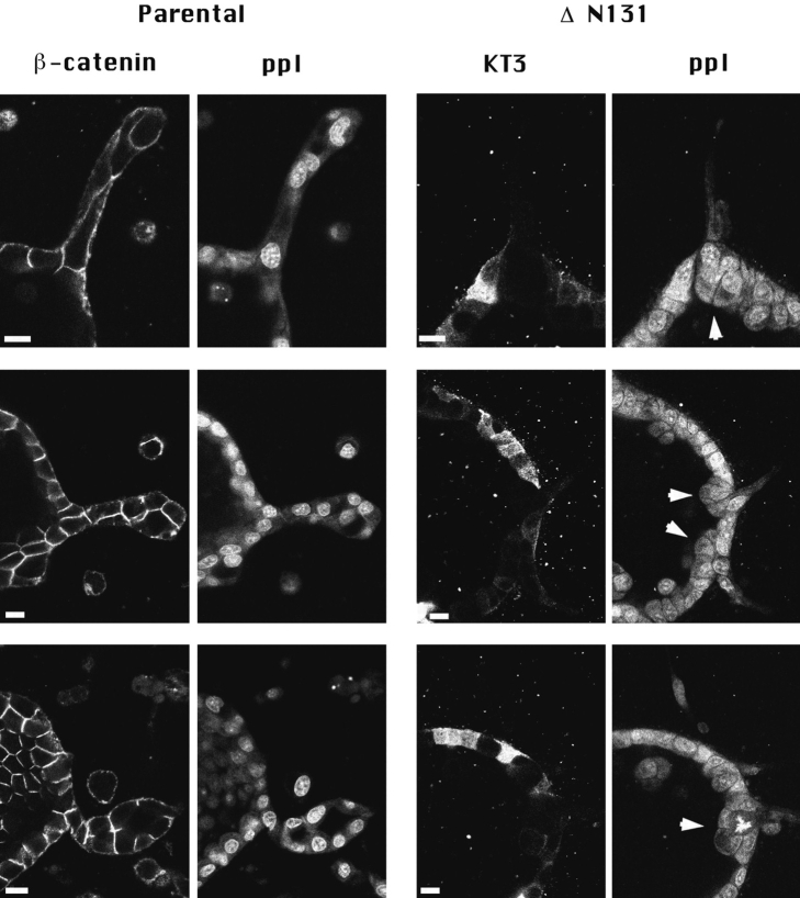
Epithelial tubulogenesis involves complex cell rearrangements that require control of both cell adhesion and migration, but the molecular mechanisms regulating these processes during tubule development are not well understood. Interactions of the cytoplasmic protein, beta-catenin, with several molecular partners have been shown to be important for cell signaling and cell-cell adhesion. To examine if beta-catenin has a role in tubulogenesis, we tested the effect of expressing NH2-terminal deleted beta-catenins in an MDCK epithelial cell model for tubulogenesis. After one day of treatment, hepatocyte growth factor/scatter factor (HGF/ SF)-stimulated MDCK cysts initiated tubulogenesis by forming many long cell extensions. Expression of NH2-terminal deleted beta-catenins inhibited formation of these cell extensions. Both DeltaN90 beta-catenin, which binds to alpha-catenin, and DeltaN131 beta-catenin, which does not bind to alpha-catenin, inhibited formation of cell extensions and tubule development, indicating that a function of beta-catenin distinct from its role in cadherin-mediated cell-cell adhesion is important for tubulogenesis. In cell extensions from parental cysts, adenomatous polyposis coli (APC) protein was localized in linear arrays and in punctate clusters at the tips of extensions. Inhibition of cell extension formation correlated with the colocalization and accumulation of NH2-terminal deleted beta-catenin in APC protein clusters and the absence of linear arrays of APC protein. Continued HGF/ SF treatment of parental cell MDCK cysts resulted in cell proliferation and reorganization of cell extensions into multicellular tubules. Similar HGF/SF treatment of cysts derived from cells expressing NH2-terminal deleted beta-catenins resulted in cells that proliferated but formed cell aggregates (polyps) within the cyst rather than tubules. Our results demonstrate an unexpected role for beta-catenin in cell migration and indicate that dynamic beta-catenin-APC protein interactions are critical for regulating cell migration during epithelial tubulogenesis.


Figures

Figure 1
Figure 1
Tubulogenesis is not affected by Dox. Cysts derived from the parental (T23) clone of MDCK cells were grown and treated with HGF/SF in the presence (+ Dox) or absence (− Dox) of doxycycline. Cultures grown (0 d) and treated for either 1 or 4 d with HGF/SF demonstrate that Dox had no effect on cyst formation or tubule development. Bar, 50 μm.
Figure 2
Figure 2
Expression of NH2-terminal deleted β-catenin inhibits early stages of tubulogenesis. Cysts were grown in the presence or absence of Dox from T23 (Parental) MDCK cells or from T23 cells transfected with Dox- repressible expression vectors for Lu or mutant β-catenins that contain NH2-terminal deletions of 90 (ΔN90) or 131 (ΔN131) amino acids. Nomarski interference contrast microscopy images of cysts treated 1 d with HGF/SF are shown. Top images (+ Dox) show typical examples of cysts in which expression of transfected proteins was inhibited with Dox. Bottom images (− Dox) show cysts in which Lu, ΔN90, or ΔN131 are expressed. Note that cysts expressing ΔN90 or ΔN131 β-catenins have fewer extensions. Representative images are shown. Experiments were repeated two to four times for each clone. Bar, 50 μm.
Figure 3
Figure 3
Quantification of tubulogenetic response after 1 d of treatment with HGF/SF. Cysts derived from T23 (Parental) MDCK cells, clone β-cat*–10 expressing full length β-catenin, two different luciferase clones (Lu-C and Lu-D), two different ΔN90 clones (ΔN90-A and -2), and three different clones of ΔN131 (ΔN131-D, -5, -7) were grown and treated for 1 d in the presence or absence of Dox. The number of extensions per cyst for each clone, ±Dox were counted in the midline focal plane of each cyst. Histograms in A show binned numbers of extensions per cyst. A representative experiment for each clone is shown. n, total number of cysts counted for each clone and experimental condition. The graph in B shows the average number of extensions per cyst (mean ± SEM). Quantification was obtained from two experiments for each clone. ▨ , + Dox; ▪, − Dox.
Figure 3
Figure 3
Quantification of tubulogenetic response after 1 d of treatment with HGF/SF. Cysts derived from T23 (Parental) MDCK cells, clone β-cat*–10 expressing full length β-catenin, two different luciferase clones (Lu-C and Lu-D), two different ΔN90 clones (ΔN90-A and -2), and three different clones of ΔN131 (ΔN131-D, -5, -7) were grown and treated for 1 d in the presence or absence of Dox. The number of extensions per cyst for each clone, ±Dox were counted in the midline focal plane of each cyst. Histograms in A show binned numbers of extensions per cyst. A representative experiment for each clone is shown. n, total number of cysts counted for each clone and experimental condition. The graph in B shows the average number of extensions per cyst (mean ± SEM). Quantification was obtained from two experiments for each clone. ▨ , + Dox; ▪, − Dox.
Figure 4
Figure 4
Expression of ΔN90 or ΔN131 β-catenin inhibits formation of cell extensions. Cysts derived from ΔN90, ΔN131, and luciferase clones were grown and treated for 1 d with HGF/SF in the presence (A, F, and K) or absence (B–E, G–J, and L–O) of Dox and processed for double immunofluorescence labeling of KT3 and ppI (A– C′ and F–H′) or anti-luciferase (anti-Lu) and ppI (K–M′), KT3 alone (D, E, I, and J), or anti-Lu alone (N and O). In A, F, and K, the weak KT3 and anti-Lu staining shows that Dox inhibits expression of mutant β-catenins and luciferase. ppI staining in A′, F′, and K′ shows that cysts from all clones treated with HGF/SF plus Dox formed multiple extensions. Cultures in B–J and L–O were treated with HGF/ SF minus Dox. Staining with KT3 or anti-Lu detects cells with high (closed arrowheads) and low (open arrowheads) ΔN90, ΔN131, and luciferase expression. (B–J) Cells expressing high amounts of mutant β-catenins do not form extensions. Extensions formed from cells with low mutant β-catenin expression are detected with ppI. Subcellular clusters of mutant β-catenins are shown at tips of cells adjacent to extending cells (D and G; closed arrows) and at tips of cell extensions (D and I; open arrows). (L–O) Cells with both high and low luciferase expression form extensions. Bars: (A, E, F, J–O) 20 μm; (B–D and G–I) 10 μm.
Figure 5
Figure 5
ΔN90 and ΔN131 β-catenins accumulate in clusters with APC protein. Parental cell cysts (A, A′, A′′, B, and C) and cysts expressing either ΔN90 (D, D′, D′′) or ΔN131 (E, E′, E′′) β-catenin were treated for 1 d with HGF/SF. Cultures were double labeled for APC protein (A′, D′, and E′) and either endogenous β-catenin (A) or KT3 (D and E). Merged images are shown in A′′, B, C, D′′, and E′′. Extensions from parental cysts contained bright, linear staining of APC (A′, open arrows) that did not colocalize with endogenous β-catenin (A′, B, C). Punctate APC protein staining is sometimes detected at tips of parental cell extensions (B and C, closed arrowheads) and occasionally colocalizes with endogenous β-catenin (A, open arrowheads). Punctate clusters of KT3 staining (D and E, closed arrows) are strikingly detected at tips of extensions of cells expressing ΔN90 and ΔN131 β-catenin. Punctate clusters of APC protein detected in D′ and E′ (closed arrowheads) colocalize with clusters of mutant β-catenin staining (D′′ and E′′, open arrowheads). Bar, 10 μm.
Figure 6
Figure 6
Stimulation of tubulogenesis from cysts, in which cell extension is inhibited, results in the formation of cell aggregates (polyps). Cysts derived from Parental (left column) and ΔN131 β-catenin clones (right column) were stimulated for 4 d with HGF/SF in the absence of Dox. Double immunofluorescence images of endogenous β-catenin and ppI or KT3 and ppI show that cells accumulate in aggregates at the base of short cell extensions formed from cells expressing low levels of ΔN131 β-catenin (arrowheads) but not at the base of tubules developing from parental cysts. Bars, 10 μm.

Similar articles

Cited by

References

    1. Aberle H, Butz S, Stappert J, Weissig H, Kemler R, Hoschuetzky H. Assembly of the cadherin-catenin complex in vitro with recombinant proteins. J Cell Sci. 1994;107:3655–3663. - PubMed
    1. Barth AIM, Pollack AL, Altschuler Y, Mostov KE, Nelson WJ. NH2-terminal deletion of β-catenin results in stable colocalization of mutant β-catenin with APC protein and altered MDCK cell adhesion. J Cell Biol. 1997;136:693–706. - PMC - PubMed
    1. Behrens J, von Kries JP, Kuhl M, Bruhn L, Wedlich D, Grosschedl R, Birchmeier W. Functional interaction of β-catenin with the transcription factor LEF-1. Nature (Lond) 1996;382:638–642. - PubMed
    1. Coleman S, Silberstein GB, Daniel CW. Ductal morphogenesis in the mouse mammary gland: evidence supporting a role for epidermal growth factor. Dev Biol. 1988;127:304–315. - PubMed
    1. Cox RT, Kirkpatrick C, Peifer M. Armadillo is required for adherens junction assembly, cell polarity, and morphogenesis during Drosophilaembryogenesis. J Cell Biol. 1996;134:133–148. - PMC - PubMed

Publication types