. 1997 Jul 15;17(14):5297-304.
doi: 10.1523/JNEUROSCI.17-14-05297.1997.

ATP P2X receptors mediate fast synaptic transmission in the dorsal horn of the rat spinal cord

Affiliations

ATP P2X receptors mediate fast synaptic transmission in the dorsal horn of the rat spinal cord

R Bardoni et al. J Neurosci. .

Abstract

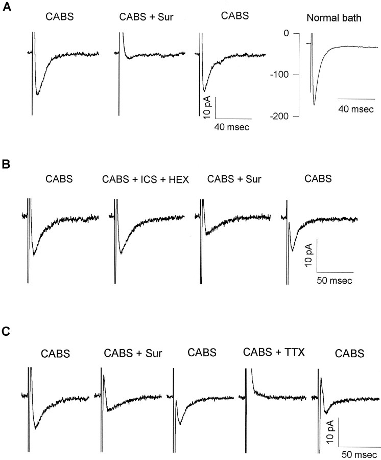
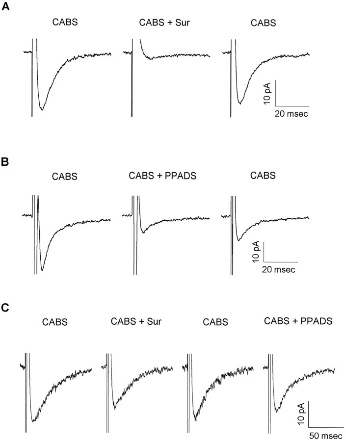
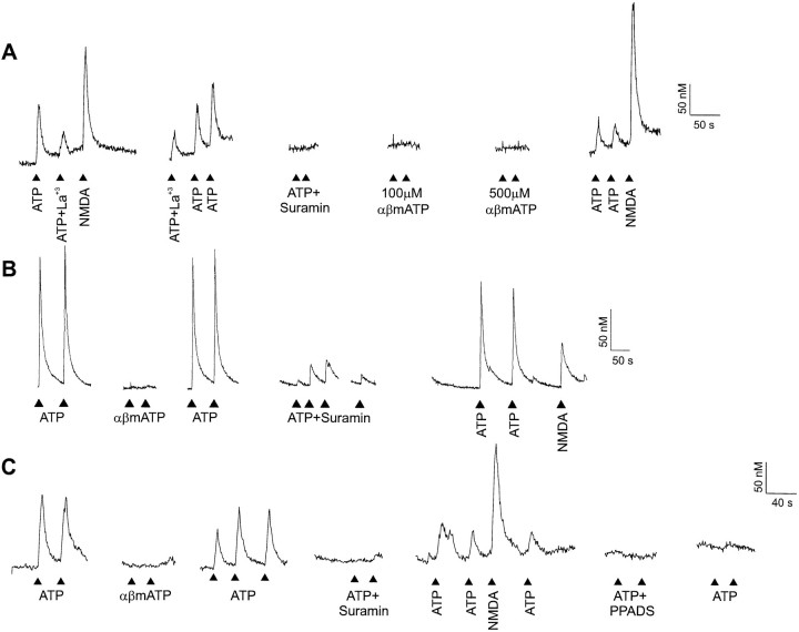
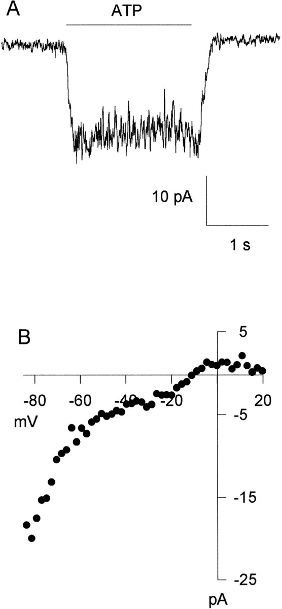
ATP has been proposed to mediate synaptic transmission in the spinal cord dorsal horn, particularly in the pathway carrying nociceptive information. Using transverse spinal cord slices from postnatal rats, we show that EPSCs mediated by P2X receptors, and presumably activated by synaptically released ATP, are evoked in a subpopulation of spinal cord lamina II neurons, a region known to receive strong input from nociceptive primary afferents. The P2X receptors on acutely dissociated dorsal horn neurons are nondesensitizing, insensitive to alphabeta methylene ATP, and show strong but variable sensitivity to the antagonists suramin and pyridoxal-phosphate-6-azophenyl-2',4'-disulfonic acid (PPADS). These characteristics are consistent with a heterogeneous population of P2X receptors, the composition of which includes P2X2, P2X4, and P2X6 receptor subtypes. Our results suggest that ATP-activated P2X receptors in lamina II of the rat spinal cord may play a role in transmitting or modulating nociceptive information.


Figures

Fig. 1.
Fig. 1.
The CABS-insensitive EPSC is mediated by P2 receptors. A, EPSCs not blocked by the “antagonist cocktail” (CABS; 10 μmCNQX, 50 μm APV, 10 μm bicuculline, and 5 μm strychnine to block AMPA, NMDA, GABAA, and glycine receptors, respectively) were blocked by suramin. EPSCs were evoked in a lamina II neuron from a P7 rat in CABS. They were abolished completely by 500 μmsuramin (CABS + Sur). After suramin washout, the CABS-insensitive synaptic current recovered. In normal Krebs’ bath (Normal bath), a larger EPSC was revealed. All traces are averages of five consecutive synaptic responses. B, EPSCs recorded from a lamina II neuron (P11) were resistant to all non-P2X receptor antagonists tested. EPSCs were recorded in CABS as the control solution. Bath application of 100 μmhexamethonium and 1 μm ICS 209–930 (CABS + ICS + HEX) did not affect EPSCs, indicating that the CABS-insensitive EPSCs were not mediated by either nicotinic acetylcholine or 5-HT3 receptors. EPSCs were partially blocked by 500 μm suramin (CABS + Sur). The suramin block was reversible, and almost complete recovery was obtained after a 15 min washout. EPSCs are averages of 10 consecutive synaptic responses. C, TTX blocked the suramin-sensitive EPSC. CABS-insensitive-evoked EPSCs recorded in a lamina II neuron (P10) were blocked by 100 μm suramin (CABS + Sur). The EPSC partially recovered after suramin washout. The suramin-sensitive EPSC was blocked in the presence of 0.5 μm TTX (CABS + TTX), indicating that ATP was released from presynaptic terminals. EPSC partially recovered after washout of TTX. EPSCs are averages of 20 consecutive synaptic responses. CABS-insensitive data for all three cells are shown with holding currents subtracted. In all cases, holding current varied <10 pA throughout the recording period.
Fig. 2.
Fig. 2.
P2X receptor antagonists variably block the P2X receptor-mediated EPSCs in lamina II neurons.A, ATP-mediated EPSCs were tested for the degree of suramin block. Fast EPSCs were recorded from a P10 neuron in CABS. Application of 500 μm suramin (CABS + Sur) completely blocked the EPSC. A complete recovery was observed after suramin washout. EPSCs are averages of 10 consecutive traces.B, ATP-mediated EPSCs were tested for the degree of PPADS block. EPSCs were recorded from a lamina II neuron (P6) in CABS. Bath application of 100 μm PPADS (CABS + PPADS) incompletely blocked the EPSC. Partial recovery was obtained after a 10 min wash in CABS. EPSCs are averages of 13–15 consecutive traces. C, P2X receptor-mediated EPSCs were tested for sensitivity to both suramin and PPADS. EPSCs were evoked in a P11 lamina II neuron in the presence of CABS. Suramin (500 μm) (CABS + Sur) partially blocked the EPSC. After a wash in CABS the suramin-blocked component recovered. Application of 50 μm PPADS (CABS + PPADS) slightly depressed the EPSC amplitude. EPSCs are averages of 20 consecutive traces. Data for all three cells are shown with holding currents subtracted. In all cases, holding current varied <10 pA throughout the recording period.
Fig. 3.
Fig. 3.
ATP-evoked [Ca2+]i transients are mediated by P2X receptors expressed on acutely dissociated dorsal horn neurons. A, ATP caused increases in [Ca2+]i in acutely dissociated dorsal horn neurons. ATP (100 μm) increased [Ca2+]i in a neuron from a P8 animal. This response was blocked completely by 100 μm suramin and partially reduced by 30 μmLa3+, a voltage-gated Ca2+ channel blocker. αβ Methylene (100 and 500 μm) ATP did not evoke an increase in [Ca2+]i. Neuronal response was confirmed by application of 100 μm NMDA.B, The response to ATP in this neuron (P10) exhibited incomplete block by suramin. C, In a dorsal horn neuron from a P7 rat, 50 μm PPADS irreversibly blocked responses to ATP.
Fig. 4.
Fig. 4.
ATP evokes nondesensitizing whole-cell currents in acutely dissociated dorsal horn neurons. A, A 2 sec application of ATP evoked a small, nondesensitizing, inward current in a neuron obtained from a P8 animal. B, The ATP-evoked current shows inward rectification in a different neuron from the same preparation. The current–voltage curve for the ATP-evoked current was constructed by applying a voltage ramp during the agonist application. Inward rectification of theI–V relationship is evident over the voltage range between −85 and −20 mV.

References

    1. Bardoni R, Goldstein PA, Lee CJ, MacDermott AB. Patch-clamp recording from visually identified substantia gelatinosa neurons in postnatal rat spinal cord slices. Soc Neurosci Abstr. 1995;21:613.
    1. Benham CD, Tsien RW. A novel receptor-operated Ca2+-permeable channel activated by ATP in smooth muscle. Nature. 1987;328:275–278. - PubMed
    1. Brake AJ, Wagenbach MJ, Julius D. New structural motif for ligand-gated ion channels defined by an ionotropic ATP receptor. Nature. 1994;371:519–523. - PubMed
    1. Buell G, Lewis C, Collo G, North RA, Surprenant A. An antagonist-insensitive P2X receptor expressed in epithelia and brain. EMBO J. 1996;15:55–62. - PMC - PubMed
    1. Burnstock G, Dumsday B, Smythe A. Atropine-resistant excitation of the urinary bladder: the possibility of transmission via nerves releasing a purine nucleotide. Br J Pharmacol. 1972;44:451–461. - PMC - PubMed

Publication types

Substances

LinkOut - more resources