. 1997 Jul 15;17(14):5380-94.
doi: 10.1523/JNEUROSCI.17-14-05380.1997.

A novel type of GABAergic interneuron connecting the input and the output regions of the hippocampus

Affiliations

A novel type of GABAergic interneuron connecting the input and the output regions of the hippocampus

K Ceranik et al. J Neurosci. .

Abstract

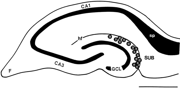
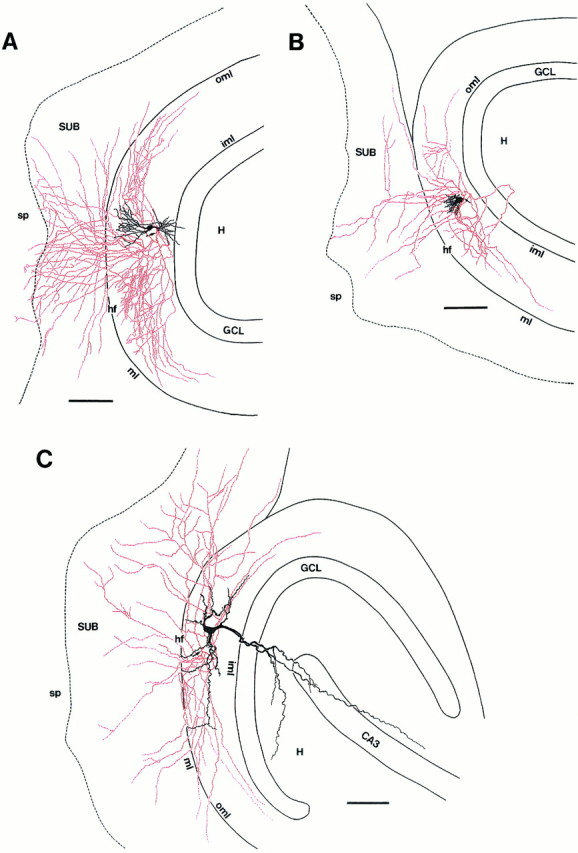
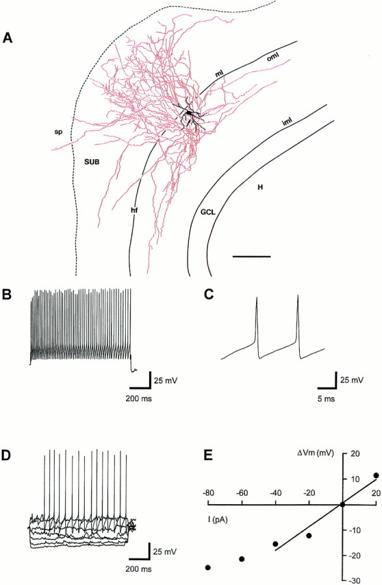
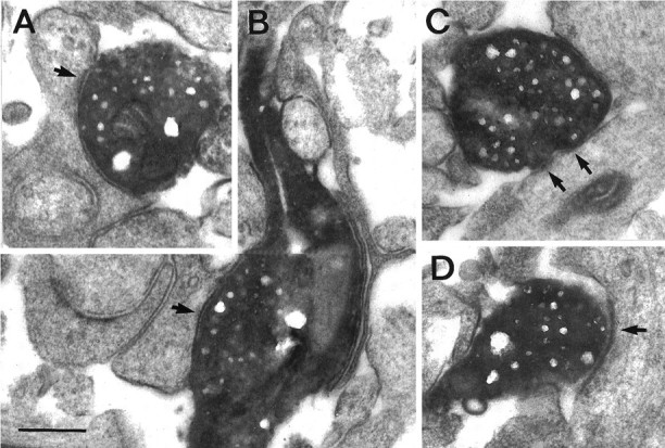
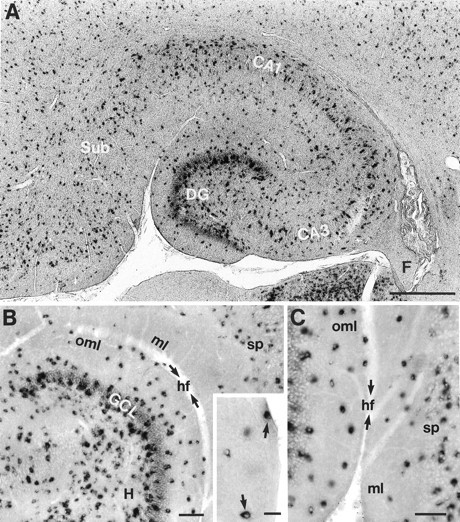
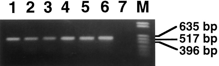
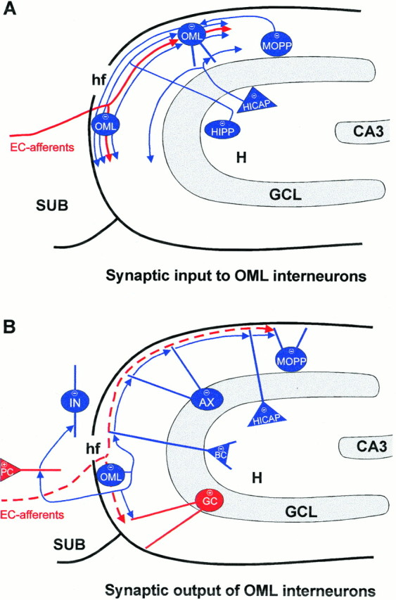
The main excitatory pathway of the hippocampal formation is controlled by a network of morphologically distinct populations of GABAergic interneurons. Here we describe a novel type of GABAergic interneuron located in the outer molecular layer (OML) of the rat dentate gyrus with a long-range forward projection from the dentate gyrus to the subiculum across the hippocampal fissure. OML interneurons were recorded in hippocampal slices by using the whole-cell patch-clamp configuration. During recording, cells were filled with biocytin for subsequent light and electron microscopic analysis. Neurons projecting to the subiculum were distributed throughout the entire OML. They had round or ovoid somata and a multipolar dendritic morphology. Two axonal domains could be distinguished: an extensive, tangential distribution within the OML and a long-range vertical and tangential projection to layer 1 and stratum pyramidale of the subiculum. Symmetric synaptic contacts were established by these interneurons on dendritic shafts in the OML and subiculum. OML interneurons were characterized physiologically by short action potential duration and marked afterhyperpolarization that followed the spike. On sustained current injection, they generated high-frequency (up to 130 Hz, 34 degrees C) trains of action potentials with only little adaptation. In situ hybridization and single-cell RT-PCR analysis for GAD67 mRNA confirmed the GABAergic nature of OML interneurons. GABAergic interneurons in the OML projecting to the subiculum connect the input and output regions of the hippocampus. Hence, they could mediate long-range feed-forward inhibition and may participate in an oscillating cross-regional interneuron network that may synchronize the activity of spatially distributed principal neurons in the dentate gyrus and the subiculum.


Figures

Fig. 1.
Fig. 1.
Location of somata of interneurons in the OML projecting to the subiculum. Schematic drawing of a transverse hippocampal slice shows the distribution of recorded and anatomically analyzed neurons. Internal reference numbers are shown within circles. Because of the overlap in the position of the somata, two neurons were omitted. Note that subiculum-projecting neurons could be found throughout the entire OML. CA1,CA3, Hippocampal regions CA1 and CA3; F, fimbria; GCL, granule cell layer; hf, hippocampal fissure; sp, stratum pyramidale of the subiculum; SUB, subiculum. Scale bar, 1 mm.
Fig. 2.
Fig. 2.
Morphology of biocytin-filled neurons in the OML. Three representative examples of OML interneurons are illustrated showing dendritic variability. A, Interneuron recorded and filled in a postnatal day 17 (P17) hippocampal slice with a fusiform morphology. The soma is located in the middle third of the OML. B, Multipolar interneuron from a P13 rat situated near the hippocampal fissure. The very short thin dendrites are confined to the OML. The main axon is marked by the open arrow, and projecting collaterals are marked by filled arrows. C, Pyramidal-like interneuron recorded and filled at P11, located in the inner third of the OML. The long apical dendrite descends into the hilar region via the granule cell layer. D, Single fiber (arrows) crossing the hippocampal fissure (hf).E, Photo montage of a displaced granule cell recorded and filled at P17. The soma is located in the inner third of the OML, and the main axon (open arrow) descends through the granule cell layer, giving rise to several collaterals (filled arrows) within the hilar region. In all micrographs the hippocampal fissure (hf) is delineated by the dashed line. GCL, Granule cell layer; H, hilar region; iml, inner molecular layer of the dentate gyrus; ml, molecular layer of the subiculum; oml, outer molecular layer of the dentate gyrus. Scale bars: in A–C, E, 50 μm; in D, 25 μm.
Fig. 3.
Fig. 3.
Axonal and dendritic morphology of three subiculum-projecting interneurons in the OML. Somata and dendrites of the neurons are drawn in black; axons are drawn inred. Arrows point to the origin of the axons. A, Camera lucida reconstruction of an interneuron (P17) with its soma located in the inner to middle third of the OML with a fusiform dendritic configuration. Note the extensive axonal arborization within layer 1 of the subiculum with terminations in stratum pyramidale and the extensive tangential spread within the OML.B, Interneuron (P13) with its soma located in the inner third of the OML with short, varicose dendrites emerging from the upper pole and restricted to the molecular layer. C, Pyramidal-like interneuron (P11) with a soma located in the inner third of the OML. This is the same neuron as that shown Figure2C. Note that the main dendrite projects through the granule cell layer into the hilar region. Neurons shown inB and C have a less extensive axonal domain within the OML and the subiculum than the cell inA. The dashed line separates layer 1 and stratum pyramidale of the subiculum. CA3, Hippocampal region CA3; GCL, granule cell layer; H, hilar region; hf, hippocampal fissure;iml, inner molecular layer of the dentate gyrus;ml, molecular layer of the subiculum;oml, outer molecular layer of the dentate gyrus;sp, stratum pyramidale of the subiculum;SUB, subiculum. Scale bars in A–C, 100 μm.
Fig. 6.
Fig. 6.
Physiological properties of a subiculum-projecting interneuron in the OML. A, Camera lucida reconstruction of an interneuron recorded and filled at P18, with its soma located underneath the hippocampal fissure and with an extensive vertical axonal projection to the subiculum. Soma and dendrites are drawn inblack; the axon is drawn in red. Thearrow points to the origin of the axon. Thedashed line separates layer 1 and stratum pyramidale of the subiculum. For abbreviations, see Figure 3. Scale bar inA, 100 μm. B, High-frequency train of action potentials (55 Hz) evoked by a 1000 msec current pulse of +180 pA. C, The second and third action potential of the voltage trace shown in B at an expanded time scale.D, Voltage responses to hyper- and depolarizing current injections of −80 to +40 pA. E, Voltage–current (V–I) relation for the voltage traces shown inD. Data points from −40 to +20 pA were fit by linear regression. RN, estimated from the slope, was 465 MΩ. The resting membrane potential was −58 mV; the holding potential was −70 mV. All data were obtained from the same neuron.
Fig. 4.
Fig. 4.
Symmetric synaptic contacts of a subiculum-projecting OML interneuron of a P14 rat. A, Biocytin-filled synaptic bouton terminating on a spine neck in the OML.B, En passant synapse established on a dendritic shaft in the OML. C, D, Two examples of synapses established on dendritic shafts in the molecular layer of the subiculum. Arrows point to what appear to be postsynaptic membrane specializations. Scale bar, 0.25 μm.
Fig. 5.
Fig. 5.
Concentric ring analysis of axonal parameters of a subiculum-projecting interneuron in the OML. A, Number of axonal segments. B, Number of axonal branch points.C, Number of boutons. D, Density of boutons per 100 μm of axonal length per ring as a function of the distance from the soma. Open and filled bars represent the distribution within the OML and subiculum, respectively. The highest bins of the histograms are marked byarrows in A–C; the total numbers (A–C) and the mean ± SD (D) are given on the right. This is the same neuron as that shown in Figure6A.
Fig. 9.
Fig. 9.
Morphological characteristics and electrophysiological properties of a displaced granule cell recorded and filled at P11. A, Camera lucida reconstruction of a displaced granule cell. The soma is located in the inner third of the OML. Note the characteristic arborization of the axon typical for granule cells. For abbreviations, see Figure 3. Scale bar inA, 100 μm. B, Action potential pattern (17 Hz) evoked by a 1000 msec current pulse of +180 pA.C, Voltage responses to hyper- and depolarizing current injections of −80 to +80 pA. D, V–Irelation for the voltage traces shown in C. Data points from −60 to +20 pA were fit by linear regression.RN was 209 MΩ, and the resting and holding membrane potentials were −70 mV. All data were obtained from the same neuron.
Fig. 7.
Fig. 7.
Distribution of GAD67-positive neurons in sections of the rat hippocampal formation at P11. A, Low-power magnification of the hippocampus and subiculum showing the overall distribution of GAD67-positive neurons. B, Distribution of GAD67-positive neurons in the hilar region, the granule cell layer, and the molecular layer of the dentate gyrus at higher magnification. The inset shows GAD67-positive neurons (arrows) located close to the hippocampal fissure.C, GAD67-positive neurons in the OML and layer 1 of the subiculum. The arrows in B andC indicate the hippocampal fissure (hf). CA1, CA3, Hippocampal regions CA1 and CA3; DG, dentate gyrus;F, fimbria; GCL, granule cell layer;H, hilar region; ml, molecular layer of the subiculum; oml, outer molecular layer of the dentate gyrus; sp, stratum pyramidale of the subiculum;Sub, subiculum. Scale bars: in A, 500 μm; in B, C, 100 μm; ininset, 25 μm.
Fig. 8.
Fig. 8.
Single-cell RT-PCR of GAD67 mRNA content of interneurons in the OML. Shown is an ethidium bromide-stained gel of cDNA fragments amplified from six electrophysiologically characterized OML interneurons by using primers for GAD67 (lanes 1–6). In addition, RT-PCR for GAD67 mRNA was performed for six CA1 pyramidal neurons. An example is shown in lane 7; note the absence of any detectable PCR product.M, DNA molecular weight marker.
Fig. 10.
Fig. 10.
Schematic drawing of putative input–output connections of subiculum-projecting interneurons in the OML.A, OML interneurons may receive synaptic input from entorhinal afferents and from various GABAergic interneurons in the molecular layer and hilar region of the hippocampus. The excitatory input via the perforant path is drawn in red. Note the coalignment of entorhinal afferents and axons of OML interneurons.B, Interneurons in the OML may establish synaptic contacts with excitatory glutamatergic principal neurons (+), granule cells (GC), and pyramidal cells (PC) in the subiculum and with various GABAergic interneurons (−) in the subiculum, the dentate molecular layer, the granule cell layer, and the hilar region. AX, Axo–axonic cell; BC, basket cell; CA3, hippocampal region CA3;EC-afferents, entorhinal afferents; GCL, granule cell layer; H, hilar region; hf, hippocampal fissure; HICAP, hilar interneuron with an axonal plexus associated with the commissural/associational path;HIPP, hilar interneuron with an axonal plexus associated with the perforant path; IN, interneuron in layer 1 of the subiculum; MOPP, molecular layer interneuron with an axonal plexus associated with the perforant path; SUB, subiculum.

References

    1. Amaral DG. A Golgi study of cell types in the hilar region of the hippocampus in the rat. J Comp Neurol. 1978;182:851–914. - PubMed
    1. Amaral DG, Witter MP. Hippocampal formation. In: Paxinos G, editor. The rat nervous system, 2nd Ed. Academic; New York: 1995. pp. 443–493.
    1. Andersen P, Eccles JC, Løyning Y. Recurrent inhibition in the hippocampus with identification of the inhibitory cell and its synapses. Nature. 1963;198:540–542. - PubMed
    1. Bender R, Plaschke M, Naumann T, Wahle P, Frotscher M. Development of cholinergic and GABAergic neurons in the rat medial septum: different onset of choline acetyltransferase and glutamate decarboxylase mRNA expression. J Comp Neurol. 1996;372:204–214. - PubMed
    1. Bragin A, Jandó G, Nádasdy Z, Hetke J, Wise K, Buzsáki G. γ (40–100 Hz) oscillation in the hippocampus of the behaving rat. J Neurosci. 1995;15:47–60. - PMC - PubMed

Publication types

Substances

LinkOut - more resources