. 1997 Jul 7;186(1):39-45.
doi: 10.1084/jem.186.1.39.

Interleukin 12 (IL-12) is crucial to the development of protective immunity in mice intravenously infected with mycobacterium tuberculosis

Affiliations

Interleukin 12 (IL-12) is crucial to the development of protective immunity in mice intravenously infected with mycobacterium tuberculosis

A M Cooper et al. J Exp Med. .

Abstract

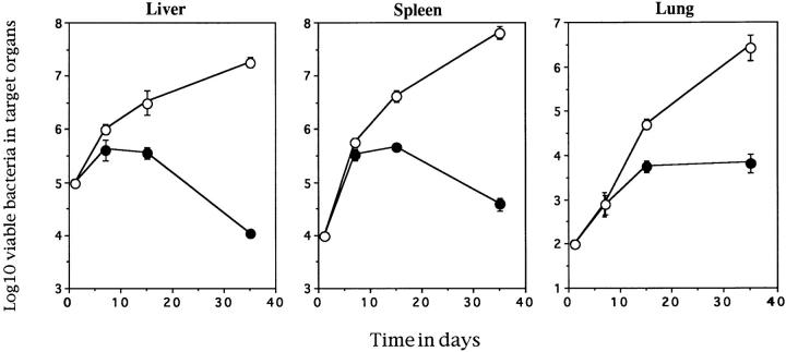
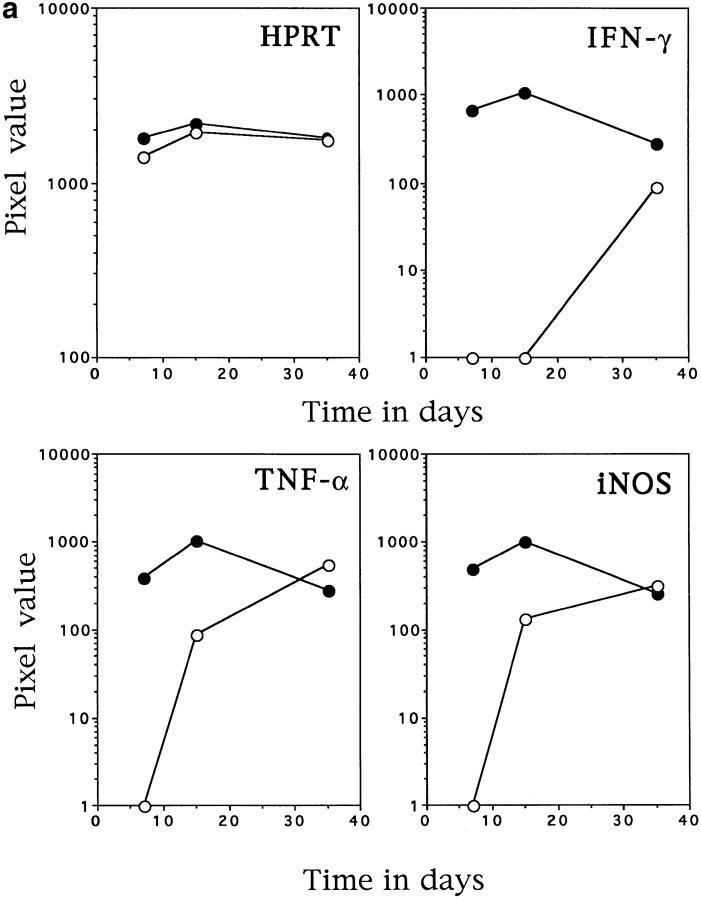
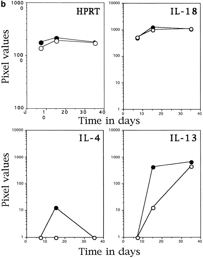
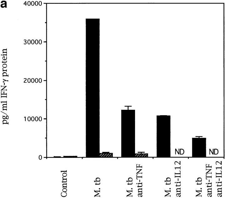
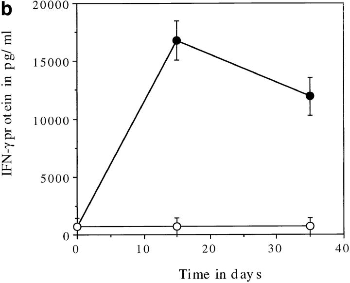
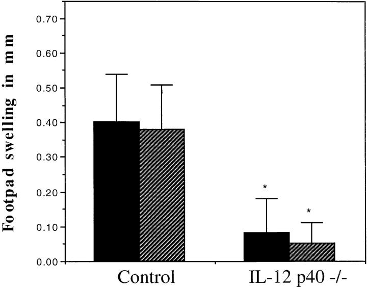
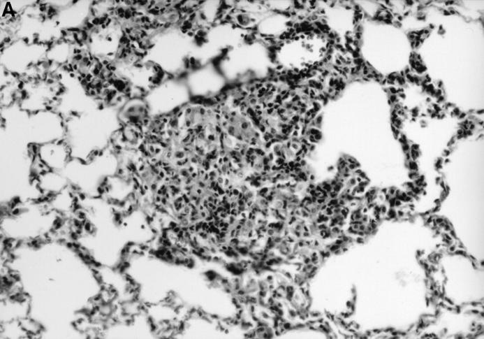
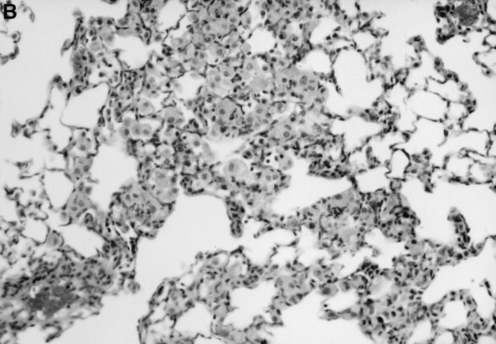
Immunity to Mycobacterium tuberculosis infection is associated with the emergence of protective CD4 T cells that secrete cytokines, resulting in activation of macrophages and the recruitment of monocytes to initiate granuloma formation. The cytokine-mediating macrophage activation is interferon-gamma (IFN-gamma), which is largely dependent on interleukin-12 (IL-12) for its induction. To address the role of IL-12 in immunity to tuberculosis, IL-12 p40(-/-) mice were infected with M. tuberculosis and their capacity to control bacterial growth and other characteristics of their immune response were determined. The IL-12 p40(-/-) mice were unable to control bacterial growth and this appeared to be linked to the absence of both innate and acquired sources of IFN-gamma. T cell activation as measured by delayed type hypersensitivity and lymphocyte accumulation at the site of infection were both markedly reduced in the IL-12 p40(-/-) mice. Therefore, IL-12 is essential to the generation of a protective immune response to M. tuberculosis, with its main functions being the induction of the expression of IFN-gamma and the activation of antigen-specific lymphocytes capable of creating a protective granuloma.


Figures

Figure 1
Figure 1
IL-12 p40−/− mice cannot control M. tuberculosis infection. Control (solid circles) and IL-12 p40−/− (open circles) mice were infected via the lateral tail vein with 105 M. tuberculosis bacteria and the number of viable bacteria present in the target organs was determined over time. The data points represent the mean and standard error of the bacterial number in the organs of four individual mice. This is a representative figure from one of two similar experiments.
Figure 2
Figure 2
IL-12 p40−/− mice have altered mRNA induction in response to infection with M. tuberculosis. Liver tissue was taken from control (solid circles) and IL-12 p40−/− (open circles) which were infected as in Fig. 1. RNA was extracted from the tissue and subjected to RT-PCR with primers specific for the molecules noted in the graphics. The HPRT graph demonstrates that equivalent readable RNA was extracted for each sample. a demonstrates the amount of IFN-γ, TNF-α, and inducible nitric oxide synthase-specific mRNA in the livers of infected mice, whereas b demonstrates the amount of IL-18, IL-4, and IL-13–specific mRNA in the same tissues. The data points represent the mean pixel values resulting from the analysis of tissue from four individual mice. This data is representative of two separate experiments.
Figure 2
Figure 2
IL-12 p40−/− mice have altered mRNA induction in response to infection with M. tuberculosis. Liver tissue was taken from control (solid circles) and IL-12 p40−/− (open circles) which were infected as in Fig. 1. RNA was extracted from the tissue and subjected to RT-PCR with primers specific for the molecules noted in the graphics. The HPRT graph demonstrates that equivalent readable RNA was extracted for each sample. a demonstrates the amount of IFN-γ, TNF-α, and inducible nitric oxide synthase-specific mRNA in the livers of infected mice, whereas b demonstrates the amount of IL-18, IL-4, and IL-13–specific mRNA in the same tissues. The data points represent the mean pixel values resulting from the analysis of tissue from four individual mice. This data is representative of two separate experiments.
Figure 3
Figure 3
IL-12 p40−/− mice cannot make IFN-γ protein in response to M. tuberculosis infection. Splenocytes from naive (a) or infected (b) mice were cultured, in vitro, at a concentration of 2 × 10⁵ cells per well in 96-well plates. In a, the cells from naive control (solid bars) or IL-12 p40−/− (striped bars) mice were exposed to live bacilli in the presence or absence of anti-cytokine antibodies (ND, not determined). In b, the cells from infected control (solid circles) or IL-12 p40−/− (open circles) mice were exposed to purified culture filtrate proteins derived from M. tuberculosis. The cells were cultured for 4 d and the supernatants were then analyzed by sandwich ELISA for IFN-γ.
Figure 3
Figure 3
IL-12 p40−/− mice cannot make IFN-γ protein in response to M. tuberculosis infection. Splenocytes from naive (a) or infected (b) mice were cultured, in vitro, at a concentration of 2 × 10⁵ cells per well in 96-well plates. In a, the cells from naive control (solid bars) or IL-12 p40−/− (striped bars) mice were exposed to live bacilli in the presence or absence of anti-cytokine antibodies (ND, not determined). In b, the cells from infected control (solid circles) or IL-12 p40−/− (open circles) mice were exposed to purified culture filtrate proteins derived from M. tuberculosis. The cells were cultured for 4 d and the supernatants were then analyzed by sandwich ELISA for IFN-γ.
Figure 4
Figure 4
IL-12 p40−/− mice fail to generate an antigen-specific recall response. Control and IL-12 p40−/− mice that were infected as in Fig. 1 were challenged in the left hind footpad with saline and the right hind footpad with 10 μg of purified protein derivative of M. tuberculosis on day 15 (solid bars) or day 30 (striped bars) of infection. The data represents the mean and standard error of the difference in footpad swelling between the saline and PPD challenged footpad for each of four mice. * P <0.05 by the Student's t test.
Figure 5
Figure 5
IL-12 p40−/− mice have defective granuloma formation. Tissue from infected control (A) and IL-12 p40−/− (B) mice was fixed in formal saline, sectioned, and stained with hematoxylin and eosin. At sites of mononuclear accumulation in the lung tissue there are more lymphocytes in the control (A) tissue compared with IL-12 p40−/− (B) tissue. Original magnification ×200.
Figure 5
Figure 5
IL-12 p40−/− mice have defective granuloma formation. Tissue from infected control (A) and IL-12 p40−/− (B) mice was fixed in formal saline, sectioned, and stained with hematoxylin and eosin. At sites of mononuclear accumulation in the lung tissue there are more lymphocytes in the control (A) tissue compared with IL-12 p40−/− (B) tissue. Original magnification ×200.

References

    1. Orme IM. The kinetics of emergence and loss of mediator T lymphocytes acquired in response to infection with Mycobacterium tuberculosis. J Immunol. 1987;138:293–298. - PubMed
    1. Orme IM, Roberts AD, Griffin JP, Abrams JS. Cytokine secretion by CD4 T lymphocytes acquired in response to Mycobacterium tuberculosis infection. J Immunol. 1993;151:518–525. - PubMed
    1. Orme IM, Miller ES, Roberts AD, Furney SK, Griffin JP, Dobos KM, Chi D, Rivoire B, Brennan PJ. T lymphocytes mediating protection and cellular cytolysis during the course of Mycobacterium tuberculosis infection. Evidence for different kinetics and recognition of a wide spectrum of protein antigens. J Immunol. 1992;148:189–196. - PubMed
    1. Orme IM. Characteristics and specificity of acquired immunologic memory to Mycobacterium tuberculosis infection. J Immunol. 1988;140:3589–3593. - PubMed
    1. Cooper AM, Dalton DK, Stewart TA, Griffin JP, Russell DG, Orme IM. Disseminated tuberculosis in interferon gamma gene-disrupted mice. J Exp Med. 1993;178:2243–2247. - PMC - PubMed

Publication types