. 1997 Jul 7;186(1):65-70.
doi: 10.1084/jem.186.1.65.

Induction of a CD8+ cytotoxic T lymphocyte response by cross-priming requires cognate CD4+ T cell help

Affiliations

Induction of a CD8+ cytotoxic T lymphocyte response by cross-priming requires cognate CD4+ T cell help

S R Bennett et al. J Exp Med. .

Abstract

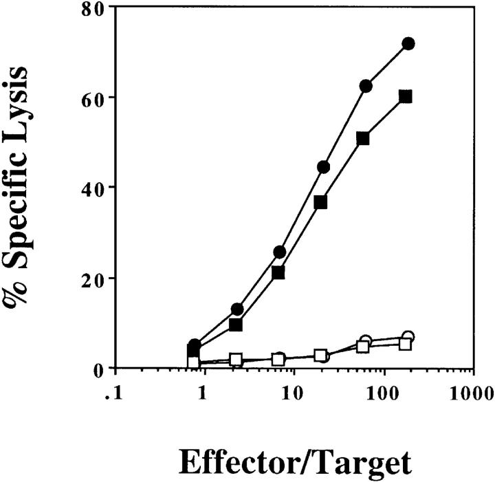
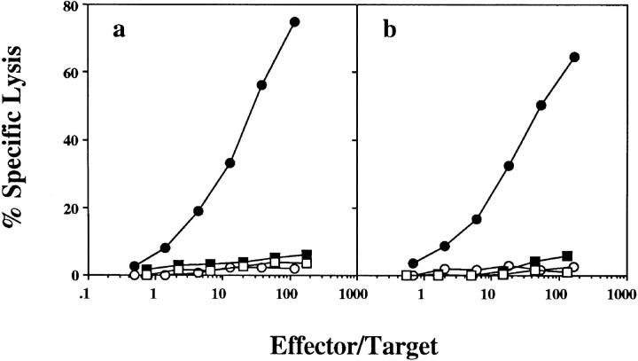
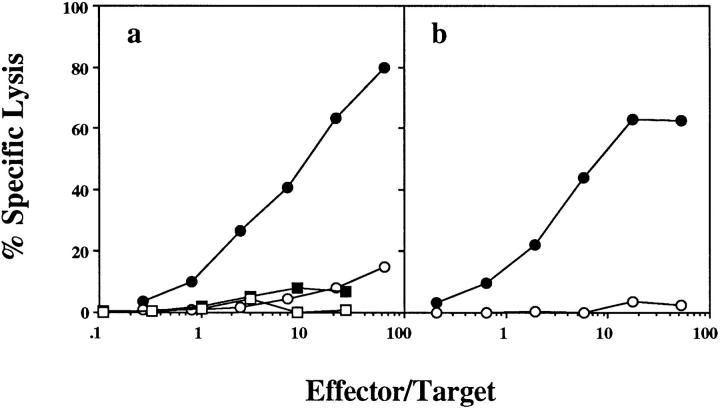
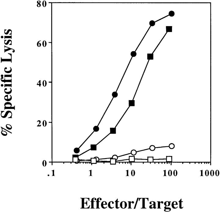
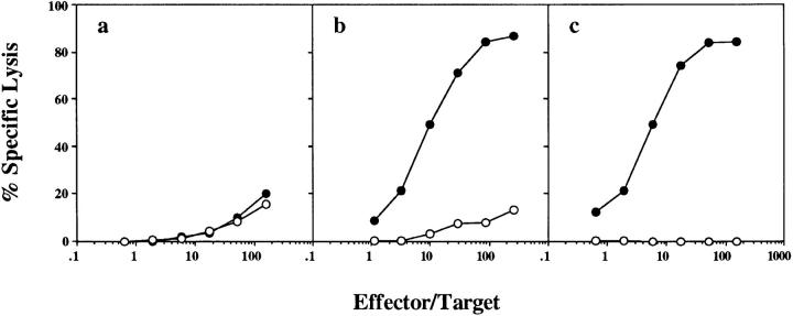
Class I-restricted presentation is usually associated with cytoplasmic degradation of cellular proteins and is often considered inaccessible to exogenous antigens. Nonetheless, certain exogenous elements can gain entry into this so-called endogenous pathway by a mechanism termed cross-presentation. This is known to be effective for class I-restricted cytotoxic T lymphocyte (CTL) cross-priming directed against a variety of exogenous tumor, viral, and minor transplantation antigens. The related effect of cross-tolerance can also effectively eliminate responses to selected self components. In both cases, this presentation appears to require the active involvement of a bone marrow-derived antigen presenting cell (APC). Here, we show that CTL induction by cross-priming with cell-associated ovalbumin requires the active involvement of CD4+ helper T cells. Importantly, this CD4+ population is only effective when both the helper and CTL determinants are recognized on the same APC. Moreover, we would argue that the cognitive nature of this event suggests that the CD4+ T cell actively modifies the APC, converting it into an effective stimulator for the successful priming of the CTL precursor.


Figures

Figure 1
Figure 1
OVA-loaded spleen cells prime by cross-presentation on host APCs. (B6 × bm1)F1 mice were injected intravenously with 2.5 × 10⁷ B6 (circles) or bm1 (squares) OVA-loaded spleen cells. 7 d later, their spleens were removed and stimulated in vitro with OVA-loaded, irradiated splenocytes. After 6 d, a chromium release assay using OVA257-264-pulsed EL4 (closed symbols) and EL4 (open symbols) targets was performed. This experiment was performed three times with 2–4 mice/group.
Figure 2
Figure 2
The host APC responsible for cross-presentation is bone marrow derived. (B6 × bm1)F1 mice were lethally irradiated, reconstituted with B6 (circles) or bm1 (squares) bone marrow and, 8 wk later, were injected intravenously with 2.5 × 10⁷ OVA-loaded bm1 (a) or B6 (b) spleen cells. 7 d later their spleens were removed and stimulated in vitro for 6 d before being tested in a chromium release assay using OVA257-264-pulsed EL4 (closed symbols) and EL4 (open symbols) targets. One of seven mice reconstituted with bm1 bone marrow and primed with B6 OVA-loaded spleen as represented in Fig. 2 b responded above background, maximizing at 26% OVA-specific lysis; no response was detected by the other six mice tested. This experiment was performed three times with 1–3 mice/group.
Figure 3
Figure 3
OVA-specific cross-priming requires CD4+ T cell help in vivo. Normal B6 mice (circles) or mice depleted of CD4+ T cells by twice weekly intraperitoneal injections of GK1.5 ascites (squares) were immunized intravenously with 2.5 × 10⁷ OVA-loaded spleen cells. 7 d later, their spleens were removed and either left intact (a) or depleted of CD4+ T cells by treatment with RL172 antibody and complement (b). After 6 d in vitro stimulation with OVA-loaded spleen cells, a CTL assay using OVA257-264-pulsed EL4 (closed symbols) and EL4 (open symbols) targets was performed. This experiment was performed four times with 2–3 mice/ group.
Figure 4
Figure 4
OVA-specific CTL induction by OVA257-264 in CFA is not dependent on CD4+ T cell help. Normal B6 mice (circles) or mice depleted of CD4+ T cells by twice weekly intraperitoneal injection of GK1.5 ascites (squares) were immunized subcutaneously with 20 μg OVA257-264 emulsified in 200 μl CFA. After 7 d, their spleens were removed and stimulated for 6 d in vitro before a chromium release assay using OVA257-264-pulsed EL4 (closed symbols) and EL4 (open symbols) targets was performed. This experiment was performed four times with 1–3 mice/group.
Figure 5
Figure 5
OVA-specific cross-presentation requires cognate CD4+ T cell help. (B6 × bm1)F1 mice were lethally irradiated and reconstituted with either equal parts of H-2A−/− (class II–deficient) and bm1 bone marrow (a and c) or (B6 × bm1)F1 bone marrow alone (b). 8 wk later, these mice were immunized with 2.0 × 10⁷ OVA-loaded bm1 spleen cells intravenously (a and b) or 10 μg OVA257-264 in 100 μl CFA subcutaneously (c). After 1 wk, their spleen cells were stimulated in vitro for 6 d and a chromium release assay using OVA257-264-pulsed EL4 (closed symbols) and EL4 (open symbols) targets was performed. This experiment was performed twice with 2–3 mice/group.
Figure 6
Figure 6
Both class II–deficient B6 and class II–sufficient bm1 bone marrow–derived APCs were present in the mixed bone marrow chimeras. Flow cytometry was performed on the peripheral blood of all mice before immunization for studies shown in Fig. 5.

References

    1. Moore MW, Carbone FR, Bevan MJ. Introduction of soluble protein into the class I pathway of antigen processing and presentation. Cell. 1988;54:777–785. - PubMed
    1. Bevan MJ. Cross-priming for a secondary cytotoxic response to minor H antigens with H-2 congenic cells which do not cross-react in the cytotoxic assay. J Exp Med. 1976;143:1283–1288. - PMC - PubMed
    1. Gooding LR, Edwards CB. H-2 antigen requirements in the in vitro induction of SV40-specific cytotoxic T lymphocytes. J Immunol. 1980;124:1258–1262. - PubMed
    1. Carbone FR, Bevan MJ. Class I–restricted processing and presentation of exogenous cell-associated antigen in vivo. J Exp Med. 1990;171:377–387. - PMC - PubMed
    1. Huang AY, Golumbek P, Ahmadzadeh M, Jaffee E, Pardoll D, Levitsky H. Role of bone marrow–derived cells in presenting MHC class I–restricted tumor antigens. Science (Wash DC) 1994;264:961–965. - PubMed

Publication types

MeSH terms

Substances