. 1997 Jul 7;186(1):109-20.
doi: 10.1084/jem.186.1.109.

Requirements for CD1d recognition by human invariant Valpha24+ CD4-CD8- T cells

Affiliations

Requirements for CD1d recognition by human invariant Valpha24+ CD4-CD8- T cells

M Exley et al. J Exp Med. .

Abstract

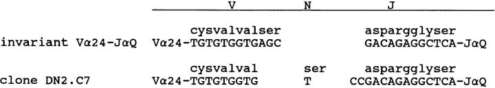
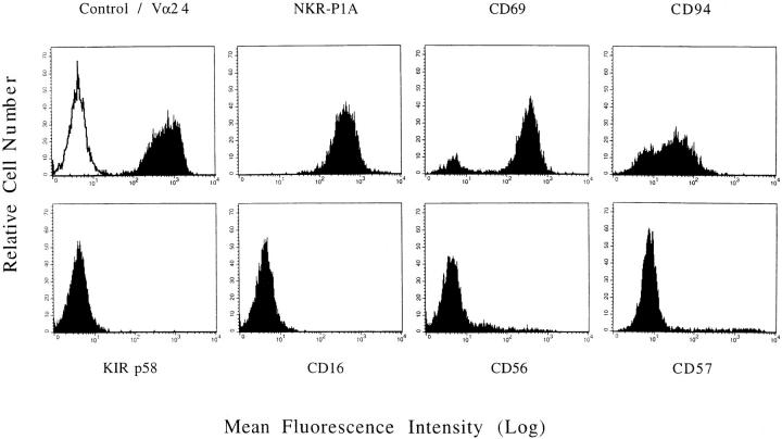
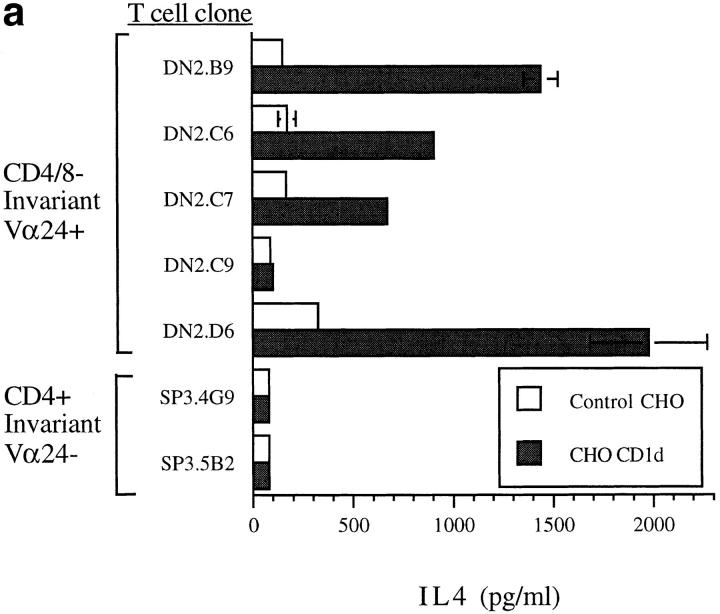
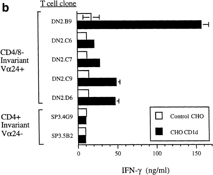
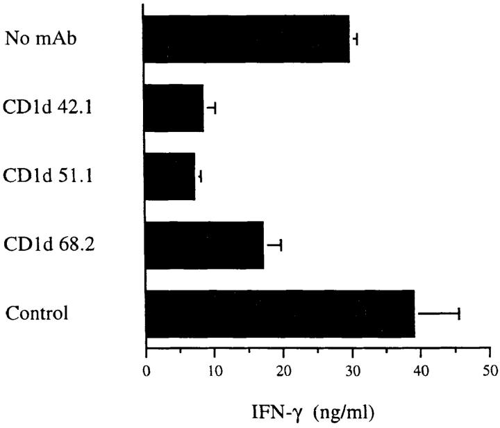
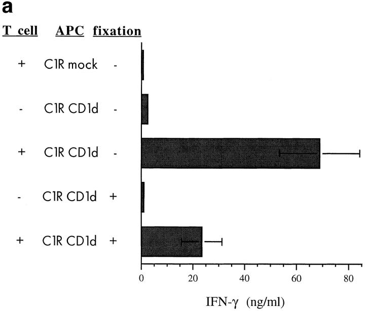
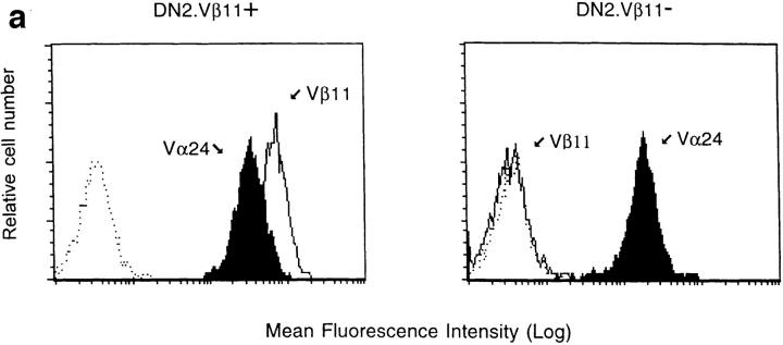
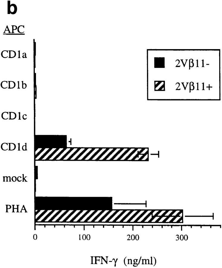
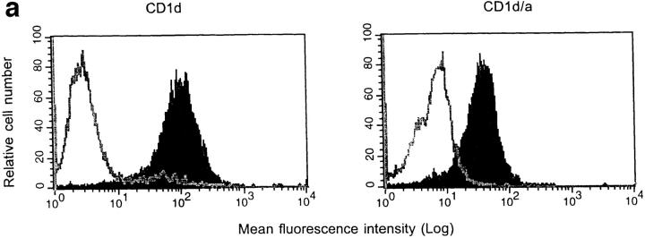
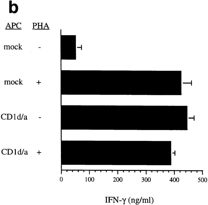
A subset of human CD4-CD8- T cells that expresses an invariant Valpha24-JalphaQ T cell receptor (TCR)-alpha chain, paired predominantly with Vbeta11, has been identified. A series of these Valpha24 Vbeta11 clones were shown to have TCR-beta CDR3 diversity and express the natural killer (NK) locus-encoded C-type lectins NKR-P1A, CD94, and CD69. However, in contrast to NK cells, they did not express killer inhibitory receptors, CD16, CD56, or CD57. All invariant Valpha24(+) clones recognized the MHC class I-like CD16 molecule and discriminated between CD1d and other closely related human CD1 proteins, indicating that recognition was TCR-mediated. Recognition was not dependent upon an endosomal targeting motif in the cytoplasmic tail of CD1d. Upon activation by anti-CD3 or CD1d, the clones produced both Th1 and Th2 cytokines. These results demonstrate that human invariant Valpha24+ CD4-CD8- T cells, and presumably the homologous murine NK1+ T cell population, are CD1d reactive and functionally distinct from NK cells. The conservation of this cell population and of the CD1d ligand across species indicates an important immunological function.


Figures

Figure 1
Figure 1
Invariant Vα24+ TCR-α nucleotide and derived amino acid sequences. DN invariant Vα24+ T cell clone cDNA was sequenced. One clone (DN2.C7) had a distinct nucleotide sequence, which resulted in an identical amino acid sequence as shown.
Figure 2
Figure 2
Expression of NK-associated proteins by invariant Vα24+ T cells. FACS® profiles of a representative DN invariant Vα24+ T cell clone (DN2.C9) 4 wk after PHA stimulation. T cells were stained with mAbs against the antigens shown or isotype-matched control mAb at 10 μg/ml and with anti-IgG FITC conjugate for 30 min each before FACS® analysis with propidium iodide gating on viable cells. (Top, left to right) P3 isotype control (open histogram) and Vα24 (solid histogram), NKR-P1A, CD69, CD94. (Bottom, left to right) p58 KIR (GL183 shown), CD16, CD56, CD57.
Figure 3
Figure 3
Invariant Vα24+ T cells responded to CD1d CHO transfectants specifically. DN invariant Vα24+ T cell clones (DN2.B9, C6, C7, C9, and D6) and control CD4+ Vα24+ invariant TCR-negative T cell clones (SP3.4G9 and 5B2), all at 2 × 10⁵/well were stimulated with 0.05% glutaraldehyde-fixed CD1d+ CHO transfectants or control CHO cells (2 × 10⁵/ well). PMA (1 ng/ml) and IL-2 (1 nM) were included, and secreted IL-4 and IFN-γ measured at 48 h by ELISA in triplicate (standard deviations shown). Similar results were obtained without IL-2. (a) IL-4; (b) IFN-γ.
Figure 3
Figure 3
Invariant Vα24+ T cells responded to CD1d CHO transfectants specifically. DN invariant Vα24+ T cell clones (DN2.B9, C6, C7, C9, and D6) and control CD4+ Vα24+ invariant TCR-negative T cell clones (SP3.4G9 and 5B2), all at 2 × 10⁵/well were stimulated with 0.05% glutaraldehyde-fixed CD1d+ CHO transfectants or control CHO cells (2 × 10⁵/ well). PMA (1 ng/ml) and IL-2 (1 nM) were included, and secreted IL-4 and IFN-γ measured at 48 h by ELISA in triplicate (standard deviations shown). Similar results were obtained without IL-2. (a) IL-4; (b) IFN-γ.
Figure 4
Figure 4
CD1d antibodies inhibited invariant Vα24+ T cell recognition of targets. DN invariant Vα24+ T cell clone DN2.D5 (105/well) was stimulated with fixed CD1d+ CHO cell transfectants (105/well) as in Fig. 3. Control and CD1d-specific mAb were included at 0.67 μg/ml, and secreted IFN-γ measured by ELISA. Higher concentrations of mAb gave similar results except for 68.2, where inhibition was more complete.
Figure 5
Figure 5
Invariant Vα24+ T cells responded to CD1d B cell transfectants. T cell clones (105/ well) were incubated for 48 h with either fixed (0.025 or 0.05% glutaraldehyde, latter shown) or unfixed C1R human B cells (105/well) transfected with CD1a, b, c, d, or plasmid alone. Results with the two fixations were indistinguishable. PMA (1 ng/ml) was included and PHA as positive control is shown for comparison. Representative IFN-γ cytokine ELISA results of multiple experiments are shown. (a) Production of IFN-γ by DN2.C9 incubated for 48 h with C1R ± fixation. (b) IFN-γ cytokine responses of three representative DN2 T cell clones incubated for 48 h with unfixed C1R transfected with CD1a, b, c, d, mock, or mock with PHA.
Figure 5
Figure 5
Invariant Vα24+ T cells responded to CD1d B cell transfectants. T cell clones (105/ well) were incubated for 48 h with either fixed (0.025 or 0.05% glutaraldehyde, latter shown) or unfixed C1R human B cells (105/well) transfected with CD1a, b, c, d, or plasmid alone. Results with the two fixations were indistinguishable. PMA (1 ng/ml) was included and PHA as positive control is shown for comparison. Representative IFN-γ cytokine ELISA results of multiple experiments are shown. (a) Production of IFN-γ by DN2.C9 incubated for 48 h with C1R ± fixation. (b) IFN-γ cytokine responses of three representative DN2 T cell clones incubated for 48 h with unfixed C1R transfected with CD1a, b, c, d, mock, or mock with PHA.
Figure 6
Figure 6
Invariant Vα24+ T cells that did not express Vβ11 responded to CD1d. (a) Representative FACS® profiles of two DN invariant TCR+ T cell lines (DN2.Vβ11+, left; and DN2.Vβ11−, right). T cells were stained with 10 μg/ml of mAbs against Vα24, Vβ11, or isotype control mAb and with anti-IgG FITC conjugate for 30 min each before FACS® analysis of viable cells. (b) T cell lines (105/well) were incubated with unfixed C1R human B cells (105/well) transfected with CD1a, b, c, d, mock, or mock with PHA as in Fig. 5 and IFN-γ production results are shown.
Figure 6
Figure 6
Invariant Vα24+ T cells that did not express Vβ11 responded to CD1d. (a) Representative FACS® profiles of two DN invariant TCR+ T cell lines (DN2.Vβ11+, left; and DN2.Vβ11−, right). T cells were stained with 10 μg/ml of mAbs against Vα24, Vβ11, or isotype control mAb and with anti-IgG FITC conjugate for 30 min each before FACS® analysis of viable cells. (b) T cell lines (105/well) were incubated with unfixed C1R human B cells (105/well) transfected with CD1a, b, c, d, mock, or mock with PHA as in Fig. 5 and IFN-γ production results are shown.
Figure 7
Figure 7
Invariant Vα24+ T cells responded to chimeric CD1d with intracellular CD1a. (a) FACS® profiles of C1R CD1d (left) and CD1d/a chimera (right) transfectants stained with normal mouse serum (open histogram) or 42.1 CD1d-specific mAb (solid histogram). (b) DN2.D6 T cell clone (105/well) was incubated with unfixed C1R human B cell CD1d/a chimera or mock transfectants (105/well) and IFN-γ cytokine ELISA results obtained.
Figure 7
Figure 7
Invariant Vα24+ T cells responded to chimeric CD1d with intracellular CD1a. (a) FACS® profiles of C1R CD1d (left) and CD1d/a chimera (right) transfectants stained with normal mouse serum (open histogram) or 42.1 CD1d-specific mAb (solid histogram). (b) DN2.D6 T cell clone (105/well) was incubated with unfixed C1R human B cell CD1d/a chimera or mock transfectants (105/well) and IFN-γ cytokine ELISA results obtained.

Similar articles

Cited by

References

    1. Martin LH, Calabi F, Lefebvre FA, Bilsland CA, Milstein C. Structure and expression of the human thymocyte antigens CD1a, CD1b, and CD1c. Proc Natl Acad Sci USA. 1987;84:9189–9193. - PMC - PubMed
    1. Aruffo A, Seed B. Expression of cDNA clones encoding the thymocyte antigens CD1a, b, c demonstrates a hierarchy of exclusion in fibroblasts. J Immunol. 1989;143:1723–1730. - PubMed
    1. Balk SP, Bleicher PA, Terhorst C. Isolation and characterization of a cDNA and gene coding for a fourth CD1 molecule. Proc Natl Acad Sci USA. 1989;86:252–256. - PMC - PubMed
    1. Blumberg RS, Gerdes D, Chott A, Porcelli SA, Balk SP. Structure and function of the CD1 family of MHC-like cell surface proteins. Immunol Rev. 1995;147:5–29. - PubMed
    1. Porcelli SA. The CD1 family: a third lineage of antigen-presenting molecules. Adv Immunol. 1995;59:1–98. - PubMed

Publication types