. 1997 Jul 7;186(1):121-9.
doi: 10.1084/jem.186.1.121.

An interleukin 5 mutant distinguishes between two functional responses in human eosinophils

Affiliations

An interleukin 5 mutant distinguishes between two functional responses in human eosinophils

M McKinnon et al. J Exp Med. .

Abstract

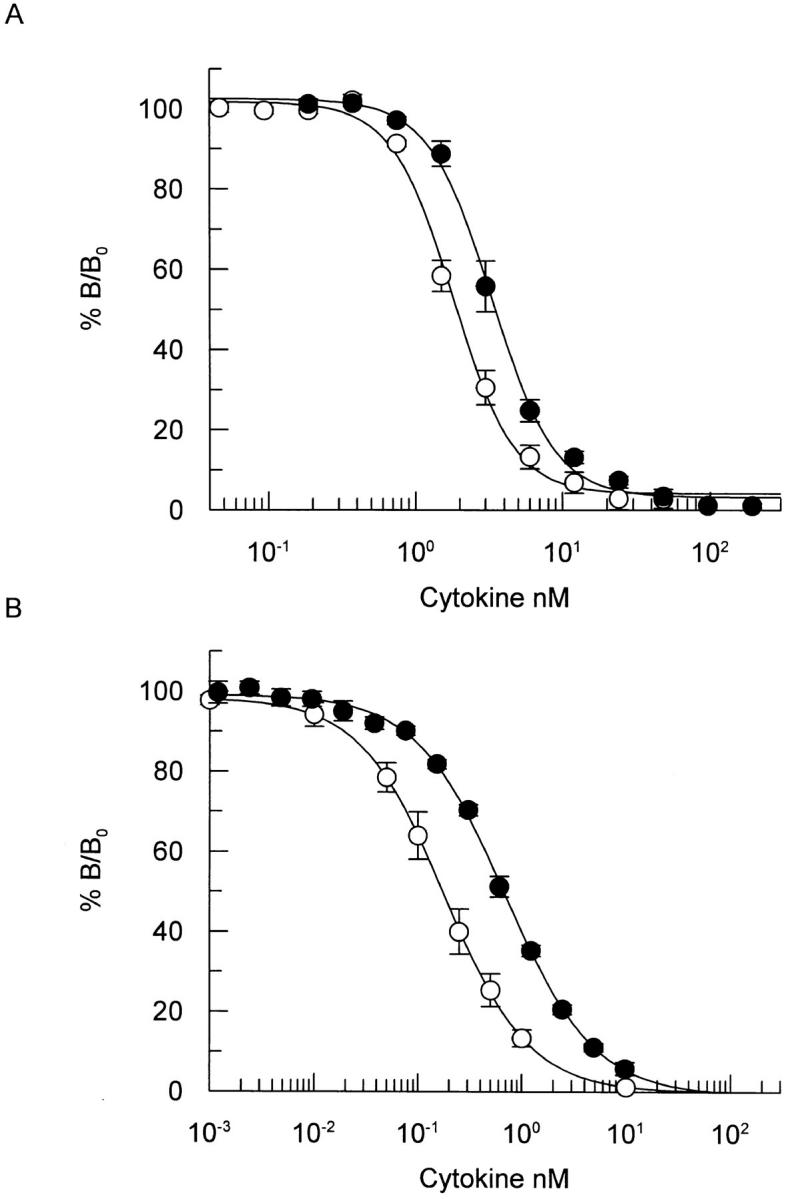
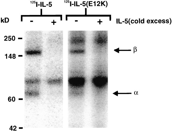
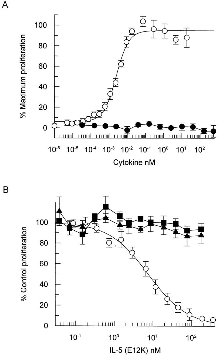
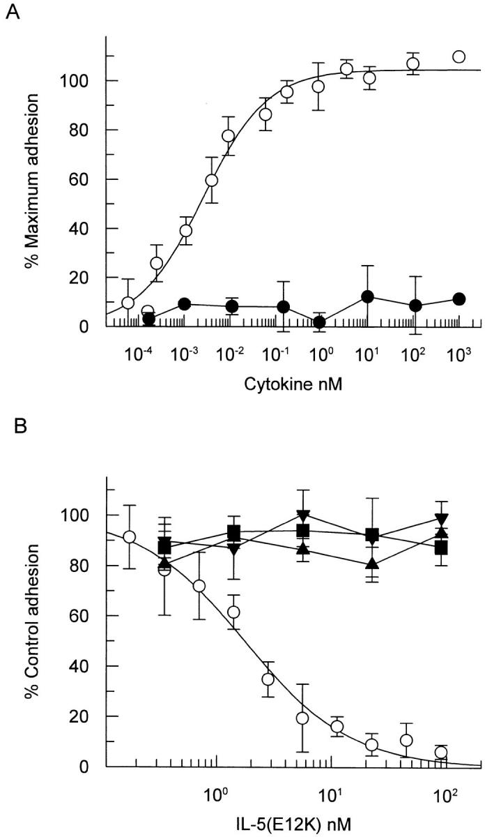
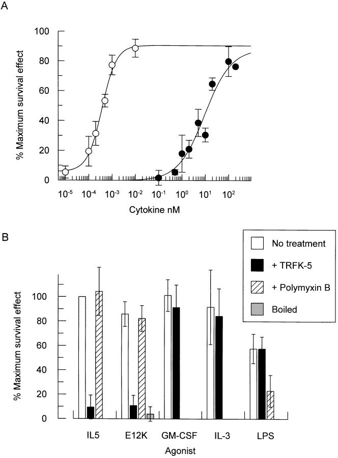
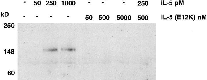
Interleukin 5 (IL-5) is the key cytokine involved in regulating the production and many of the specialized functions of mature eosinophils including priming, adhesion, and survival. We have generated a point mutant of human IL-5, IL-5 (E12K), which is devoid of agonist activity in both a TF-1 cell proliferation assay and a human eosinophil adhesion assay. However, IL-5 (E12K) is a potent and specific antagonist of both these IL-5-dependent functional responses. In both receptor binding and cross-linking studies the wild-type and IL-5 (E12K) mutant exhibit virtually identical properties. This mutant protein was unable to stimulate tyrosine phosphorylation in human eosinophils, and blocked the phosphorylation stimulated by IL-5. In contrast, IL-5 (E12K) is a full agonist in a human eosinophil survival assay, although with reduced potency compared to the wild-type protein. This IL-5 mutant enables us to clearly distinguish between two IL-5-dependent functional responses and reveals distinct mechanisms of receptor/cellular activation.


Figures

Figure 1
Figure 1
Comparative binding of wild-type IL-5 and IL-5 (E12K) to recombinant IL-5 receptor α chain (A) or to the receptor α/β complex on TF-1 cells (B). (A) Recombinant IL-5 receptor α-chain, immobilized on SPA beads, were incubated with 100 pM 125I-IL-5 in the presence of increasing concentrations of unlabeled wild-type IL-5 (open circles) or IL-5 (E12K) (closed circles). After incubation for 4 h at room temperature samples were counted in a Wallac 1450 microbeta counter set up in SPA mode. The results show competition as percentage of maximum binding (% B/Bo). Each value represents the mean ± SEM of four independent experiments for wild-type and six independent experiments for IL-5 (E12K). (B) TF-1 cells were incubated at room temperature for 2 h in the presence of 200 pM 125I-IL-5 and increasing concentrations of unlabeled wild-type (open circles) or IL-5 (E12K) (closed circles). After separation of bound and free ligand the cell-associated radioligand was quantified in a gamma counter. The results show competition as percentage of maximum binding (% B/Bo). Each value represents the mean ± SEM of five independent experiments.
Figure 2
Figure 2
Chemical cross-linking of wild-type IL-5 and IL-5 (E12K) to IL-5 receptors. COS cells transfected with human IL-5 receptor α and βc chain cDNAs were incubated with ∼70 nM 125I-IL-5 or 125I-IL-5 (E12K) for 2 h at 4°C in the presence or absence of a 100-fold molar excess of unlabeled IL-5, then cross-linked with 1 mM BS3. After detergent lysis of cells, soluble extracts were analyzed by SDS-PAGE and radiolabeled bands visualized by a PhosphoImager.
Figure 3
Figure 3
IL-5 (E12K) exhibits no agonist activity (A) and is a specific IL-5 antagonist (B) in a TF-1 cell proliferation assay. (A) TF-1 cells were incubated with increasing concentrations of either wild-type (open circles) or IL-5 (E12K) (closed circles) for 60–72 h and the induction of proliferation was measured using a nonradioactive cell proliferation assay. Each value represents the mean ± SEM of four independent experiments. (B) TF-1 cell proliferation was assayed at either 77 pM IL-5 (open circles), 133 pM IL-3 (closed squares) or 14 pM GM-CSF (closed triangles) in the presence of increasing concentrations of IL-5 (E12K). The concentrations of wild-type cytokines represent ∼ED80's (concentration of cytokine required to give 80% of the maximum biological response) for proliferation in our TF-1 cell line. Each value represents the mean ± SEM of six independent experiments. 100% values for IL-5–, IL-3–, and GM-CSF–induced proliferation were equivalent to A550 of 0.68 ± 0.02, 0.73 ± 0.18, and 0.64 ± 0.04, respectively. Unstimulated levels were 0.03 ± 0.02.
Figure 4
Figure 4
IL-5 (E12K) exhibits no agonist activity (A) and is a specific IL-5 antagonist (B) in a cytokine induced eosinophil activation assay. (A) Human eosinophils were incubated with increasing concentrations of either wild-type (open circles) or IL-5 (E12K) (closed circles) for 30 min at 37°C, in a 96-well microtiter plate precoated with human IgG. After a washing step, the adherent eosinophils were lysed and the endogenous peroxidase activity measured in a colorimetric assay. (B) In antagonist experiments eosinophils were incubated with either 20 pM IL-5 (open circles), 180 pM IL-3 (closed squares), 6 pM GM-CSF (closed triangles), or 1,000 pM TNF-α (inverted triangles) in the presence of increasing concentrations of IL-5 (E12K). The concentrations of wild-type cytokines represent ∼ED80's in the eosinophil adhesion assay. In both panels each value represents the mean ± SEM of three independent experiments using different blood donors. 100% values for IL-5–, IL-3–, GM-CSF–, and TNF-α–induced adhesion were 0.56 ± 0.07, 0.76 ± 0.05, 0.41 ± 0.07, and 0.49 ± 0.06, respectively. Unstimulated levels were 0.15 ± 0.05.
Figure 5
Figure 5
Both wild-type IL-5 and IL-5 (E12K) promote eosinophil survival (A). The agonist activity of E12K can be abolished by boiling, or pretreatment with a neutralizing anti–IL-5 antibody but not by polymyxin B (B). (A) Increasing concentrations of either wild-type (open circles) or IL-5 (E12K) (closed circles) were incubated with purified human eosinophils for 72 h at 37°C and eosinophil viability measured by trypan blue exclusion. Data expressed as a percentage of the maximum survival effect obtained with wild-type IL-5 in each experiment (66% ± 5.6 viable cells, compared to 2.7% ± 1.7 in the absence of cytokine). Each value represents the mean ± SEM of six independent experiments using different blood donors. (B) Human eosinophils were incubated with either 1 pM IL-5, 50 nM IL-5 (E12K), 40 pM IL-3, 20 pM GM-CSF, or 1 ng/ml LPS alone, or in the presence of 250 μg/ml anti–IL-5 neutralizing antibody, TRFK-5, or after pretreatment with 500 U/ml polymyxin B for 1 h at 37°C. For boiling experiments, IL-5 (E12K) was boiled for 15 min before incubation with eosinophils. Eosinophil viability was measured after 72 h by trypan blue exclusion. The concentrations of cytokines represent ∼ED80's in the eosinophil survival assay. Data expressed as a percentage of the maximum survival effect obtained with wild-type IL-5 in each experiment (61% ± 8.0 viable cells, compared to 17% ± 3.7 in the absence of cytokine). Each value represents the mean ± SEM of at least three independent experiments.
Figure 6
Figure 6
Wild-type IL-5 but not IL-5 (E12K) stimulates tyrosine phosphorylation of a 150-kD protein in human eosinophils. Human eosinophils were stimulated with the indicated concentrations of cytokines for 5 min at 37°C. Cells were pelleted, resuspended in SDS sample buffer and lysates were electrophoresed and immunoblotted with anti-phosphotyrosine antibody.

Similar articles

Cited by

References

    1. Bohjanen PR, Okajima M, Hodes RJ. Differential regulation of interleukin 4 and interleukin 5 gene expression: a comparison of T-cell gene induction by anti-CD3 antibody or by exogenous lymphokines. Proc Natl Acad Sci USA. 1990;87:5283–5287. - PMC - PubMed
    1. Plaut M, Pierce JH, Watson CJ, Hanley-Hyde J, Nordan RP, Paul WE. Mast cell lines produce lymphokines in response to cross-linkage of FcεRI or to calcium ionophores. Nature (Lond) 1989;339:64–67. - PubMed
    1. Dubucquoi S, Desreumaux P, Janin A, Klein O, Goldman M, Tavernier J, Capron A, Capron M. Interleukin 5 synthesis by eosinophils: association with granules and immunoglobulin-dependent secretion. J Exp Med. 1994;179:703–708. - PMC - PubMed
    1. Clutterbuck EJ, Hirst EM, Sanderson CJ. Human interleukin-5 (IL-5) regulates the production of eosinophils in human bone marrow cultures: comparison and interaction with IL-1, IL-3, IL-6, and GM-CSF. Blood. 1989;73:1504–1512. - PubMed
    1. Walsh GM, Hartnell A, Wardlaw AJ, Kurihara K, Sanderson CJ, Kay AB. IL-5 enhances the in vitro adhesion of human eosinophils, but not neutrophils, in a leucocyte integrin (CD11/18)-dependent manner. Immunology. 1990;71:258–265. - PMC - PubMed