. 1997 Jul 8;94(14):7176-81.
doi: 10.1073/pnas.94.14.7176.

The primary fibrin polymerization pocket: three-dimensional structure of a 30-kDa C-terminal gamma chain fragment complexed with the peptide Gly-Pro-Arg-Pro

Affiliations

The primary fibrin polymerization pocket: three-dimensional structure of a 30-kDa C-terminal gamma chain fragment complexed with the peptide Gly-Pro-Arg-Pro

K P Pratt et al. Proc Natl Acad Sci U S A. .

Abstract

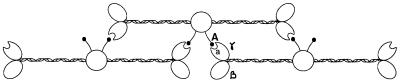
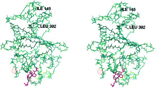
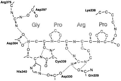
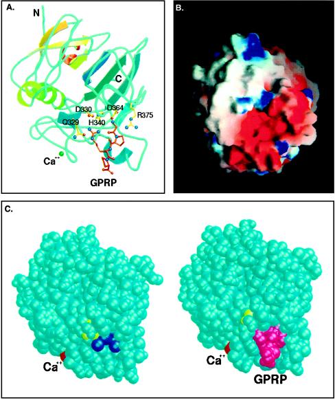
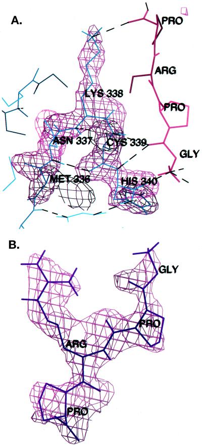
After vascular injury, a cascade of serine protease activations leads to the conversion of the soluble fibrinogen molecule into fibrin. The fibrin monomers then polymerize spontaneously and noncovalently to form a fibrin gel. The primary interaction of this polymerization reaction is between the newly exposed N-terminal Gly-Pro-Arg sequence of the alpha chain of one fibrin molecule and the C-terminal region of a gamma chain of an adjacent fibrin(ogen) molecule. In this report, the polymerization pocket has been identified by determining the crystal structure of a 30-kDa C-terminal fragment of the fibrin(ogen) gamma chain complexed with the peptide Gly-Pro-Arg-Pro. This peptide mimics the N terminus of the alpha chain of fibrin. The conformational change in the protein upon binding the peptide is subtle, with electrostatic interactions primarily mediating the association. This is consistent with biophysical experiments carried out over the last 50 years on this fundamental polymerization reaction.


Figures

Figure 1
Figure 1
Simplified model of interactions between adjacent fibrin chains at the beginning of the polymerization reaction. Thrombin cleaves the α chain N terminus, creating a new N terminus (the A site) beginning with the sequence Gly-Pro-Arg. The A site binds to the complementary “a” polymerization pocket in the γ chain during the alignment of the fibrin protofibrils.
Figure 2
Figure 2
Stereoview of the superimposed backbones of the rFbgγC30 molecules in the peptide complexed (green) and uncomplexed (purple) structures. The peptide Gly-Pro-Arg-Pro is shown in magenta, and the van der Waals surface of the calcium atom is shown as an orange dot-surface. Because the first two residues are flexible, the superposition begins with the third residue, Ile-145.
Figure 3
Figure 3
Schematic of interactions between GPRP and the protein. Hydrogen bonds and favorable ionic interactions are indicated by dotted lines. One of the terminal nitrogens of the arginine side chain is 3.26 Å away from the carbonyl oxygen of the first peptide proline, creating a weak hydrogen bond.
Figure 4
Figure 4
(A) Model of rFbgγC30 with the peptide GPRP bound at the “a” polymerization pocket. The side chains of residues Q329, D330, H340, D364, and R375 are involved in interactions that stabilize the complex with the peptide. Figure created using molscript (38) and raster3d (39). (B) End-on view of the “a” polymerization pocket, colored according to the electrostatic potential computed using the program grasp (40). The high concentration of negatively charged residues at this site, as well as the depth and narrowness of the pocket, confer its specificity for the positively charged GPR sequence of the α chain of fibrin. The model is of the peptide-complexed structure of rFbgγC30, with the peptide removed to show the binding site more clearly. (C) Space-filling models of rFbgγC30 in the uncomplexed and peptide-complexed structures. The peptide GPRP (magenta) displaces seven tightly bound water molecules (dark blue). The calcium atom is colored red, and Y363, which was shown previously by Doolittle and coworkers (7) to be close to the binding site, is colored yellow. Figure was created using molscript (38) and raster3d (39).
Figure 5
Figure 5
(A) Electron density is shown superimposed on the turn containing the cis peptide bond between K338 and C339 in the peptide-complexed structure. |Fo| − |Fc| omit maps were calculated after omitting all atoms of residues 336–340, and the map is contoured at 3σ. Hydrogen bonds are drawn as dashed lines. (B) The GPRP peptide was removed from the model and an |Fo| − |Fc| map was calculated and contoured at 3σ, then superimposed upon the peptide model.

References

    1. Davie E W, Fujikawa K, Kisiel W. Biochemistry. 1991;29:10363–10370. - PubMed
    1. Lorand L, Losowsky M S, Miloszewski K J M. Prog Hemost Thromb. 1980;5:245–290. - PubMed
    1. Hall C E, Slayter H S J. J Biophys Biochem Cytol. 1959;5:11–15. - PMC - PubMed
    1. Weisel J W, Stauffacher C V, Bullitt E, Cohen C. Science. 1985;230:1388–1391. - PubMed
    1. Gollwitzer R, Bode W, Schramm H-J, Typke D, Guckenberger R. Ann NY Acad Sci. 1983;408:214–225. - PubMed

Publication types

LinkOut - more resources