. 1997 Jul 8;94(14):7186-91.
doi: 10.1073/pnas.94.14.7186.

Interaction between replication protein A and p53 is disrupted after UV damage in a DNA repair-dependent manner

Affiliations

Interaction between replication protein A and p53 is disrupted after UV damage in a DNA repair-dependent manner

N A Abramova et al. Proc Natl Acad Sci U S A. .

Abstract

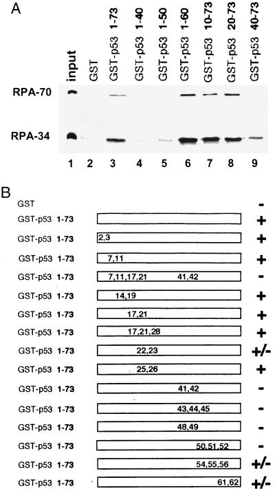
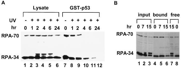
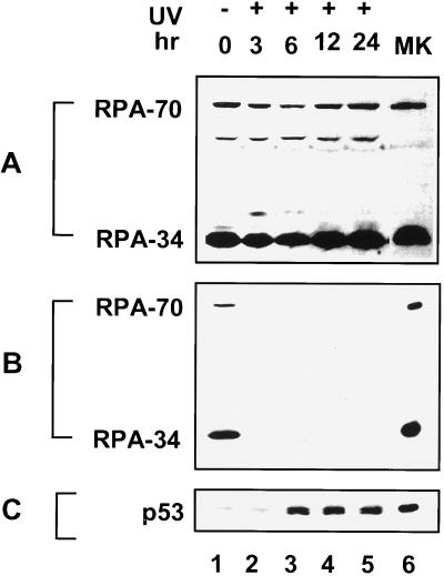
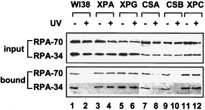
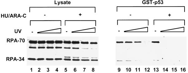
Replication protein A (RPA) is required for both DNA replication and nucleotide excision repair. Previous studies have shown that RPA interacts with the tumor suppressor p53. Herein, we have mapped a 20-amino acid region in the N-terminal part of p53 that is essential for its binding to RPA. This region is distinct from the minimal activation domain of p53 previously identified. We also demonstrate that UV radiation of cells greatly reduces the ability of RPA to bind to p53. Interestingly, damage-induced hyperphosphorylated RPA does not associate with p53. Furthermore, down-regulation of the RPA/p53 interaction is dependent upon the capability of cells to perform global genome repair. On the basis of these data, we propose that RPA may participate in the coordination of DNA repair with the p53-dependent checkpoint control by sensing UV damage and releasing p53 to activate its downstream targets.


Figures

Figure 1
Figure 1
Characterization of the RPA-interacting domain of p53. (A). Deletion analysis of the N-terminal domain of p53. One-fourth of the input purified RPA (50 ng) and 40% of the bound fractions were loaded on the gel. Approximately 25–30% of the input RPA was bound to the wild-type GST–p53 beads. (B) RPA-binding ability of various point mutants of the p53 activation domain. +, Affinity no less than the wild type; −, signal equivalent to GST alone; ±, reduced binding affinity similar to the one shown in Fig. 1A, lane 9. The positions and types of these mutations are E2K, E3K, D7H, E11K, L14Q, E17K, F19S, D21H, L22Q, W23S, L25Q, L26H, E28K, D41H, D42H, L43A, M44A, L45A, D48H, D49H, I50A, E51A, Q52A, F54A, T55A, E56A, D61H, and E62K.
Figure 2
Figure 2
RPA from UV-irradiated RKO cells loses its ability to bind the activation domain of p53. (A) Cells were irradiated at 50 J/m2 and subsequently incubated in medium at 37°C as indicated before harvesting. RPA from these lysates was tested for binding to GST–p53-(1–73). RPA was detected by immunoblotting using monoclonal antibodies raised against RPA-70 and RPA-34. The species migrating slightly above the major RPA-34 band in the lysates from UV-irradiated cells represents hyperphosphorylated RPA-34. In this and the following experiments, 5% of the input lysates (lanes 1–6) and 50% of the bound fractions (lanes 7–12) were loaded on the gel. The 0-hr point represents the sample without irradiation. (B) RPA binding to p53 is not significantly affected by 20 Gy of γ-radiation. Lysates (lanes 1–3) were harvested at different times and used in the binding assay with GST–p53 (lanes 4–6). The supernatants of the completed binding reactions at 0 and 15 hr were included (lanes 7 and 8).
Figure 3
Figure 3
UV radiation disrupts the interaction between the endogenous p53 and RPA. (A) Lysates were prepared at different times after UV irradiation at 50 J/m2. Total RPA in extracts was detected by immunoblotting using an anti-RPA polyclonal antiserum. The 50-kDa band is a degradation product of RPA-70. (B) The lysates were immunoprecipitated by an anti-p53 monoclonal antibody crosslinked to protein G-Sepharose beads. The immunoprecipitates were resolved by SDS/PAGE and immunoblotted using an anti-RPA polyclonal antiserum. (C) The same blot shown in B was stripped and reprobed with the anti-p53 antibody. Lanes 6 in A–C contain purified recombinant proteins as markers.
Figure 4
Figure 4
RPA binding to p53 is modulated by UV radiation in a NER-dependent manner. Normal human primary fibroblasts (WI38) and mutant cells with defects in various aspects of NER were irradiated at 50 J/m2. Lysates were prepared 4 hr after irradiation and tested for binding of RPA to the GST–p53 beads. RPA in the input lysates (Upper) and in the bound fractions (Lower) was detected by immunoblotting using anti-RPA-70 and anti-RPA-34 monoclonal antibodies.
Figure 5
Figure 5
RPA binding to p53 is more sensitive to UV radiation when the gap-filling synthesis of NER is inhibited. WI38 cells were irradiated with increasing doses of UV (10, 20, and 50 J/m2) and incubated for 3 hr in the absence (lanes 1–4 and 9–12) or presence of 2 mM HU and 10 μM araC (lanes 5–8 and 13–16). RPA binding to the GST–p53 beads was detected by immunoblotting with an anti-RPA polyclonal antiserum.

References

    1. Elledge S J. Science. 1996;274:1664–1672. - PubMed
    1. Paulovich A G, Toczyski D P, Hartwell L H. Cell. 1997;88:315–321. - PubMed
    1. Vogelstein B, Kinzler K W. Cell. 1992;70:523–526. - PubMed
    1. Levine A J. Cell. 1997;88:323–331. - PubMed
    1. Huang L-C, Clarkin K C, Wahl G M. Proc Natl Acad Sci USA. 1996;93:4827–4832. - PMC - PubMed

Publication types

Substances