. 1997 Jul 8;94(14):7320-5.
doi: 10.1073/pnas.94.14.7320.

Ethanolamine modulates the rate of rat hepatocyte proliferation in vitro and in vivo

Affiliations

Ethanolamine modulates the rate of rat hepatocyte proliferation in vitro and in vivo

H Sasaki et al. Proc Natl Acad Sci U S A. .

Abstract

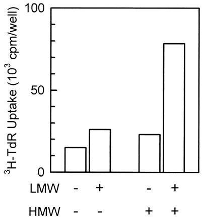
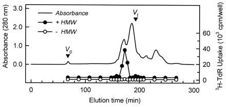
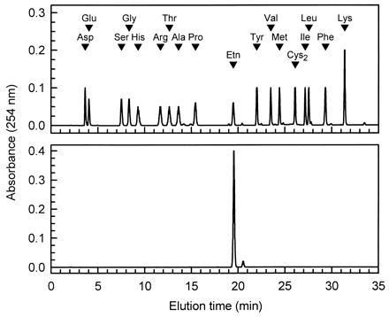
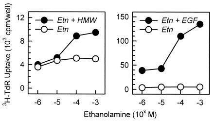
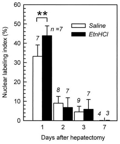
A low molecular weight, heat-resistant hepatotrophic factor in an extract from the bovine intestinal mucosa was purified and identified as ethanolamine by structural analyses. The mode of action of ethanolamine in vitro and in vivo coincided with that of the crude extract of the tissue, indicating that ethanolamine is the active component. Ethanolamine synergistically elevated the stimulation of DNA synthesis in hepatocytes in primary culture when added together with a growth factor, such as epidermal growth factor, with the ED50 being 20 microM, although it showed little stimulatory effect by itself. Contrary to these in vitro results, the intraperitoneal administration of ethanolamine hydrochloride (24 mg of ethanolamine per kg of body weight) enhanced hepatocyte proliferation in regenerating rat livers after two-thirds hepatectomy without the administration of any growth factors. In the regenerating liver, hepatocyte proliferation may be initiated by an endogenous growth factor, but the supply of ethanolamine in circulation may not be sufficient for optimal hepatocyte proliferation; thus, the exogenous administration of ethanolamine may further enhance hepatocyte proliferation. Ethanolamine in circulation may be a humoral hepatotrophic factor.


Figures

Figure 1
Figure 1
Synergistic stimulation of DNA synthesis of hepatocytes in primary culture by the LMW and HMW factors derived from the bovine small intestinal mucosa. The LMW factor (3%, vol/vol) and the HMW factor (1%, vol/vol) from the bovine small intestinal mucosa slightly stimulated DNA synthesis ([3H]thymidine uptake) when each of them was independently added to rat hepatocytes in primary culture. However, a synergistic increase in stimulation was seen when they were added simultaneously. The LMW factor and the HMW factor are referred to as LMW and HMW, respectively. All bars are the mean of duplicate assays.
Figure 2
Figure 2
Profile of gel filtration chromatography of the LMW factor. The LMW factor (10 ml) was loaded into a Sephacryl S-100 column (5 × 30 cm) equilibrated with pure water at a flow rate of 2 ml/min. Fractions (10 ml) were assayed for their activity that enhances the DNA synthesis in hepatocytes. The fractions (3%, vol/vol) were added to the culture with or without the HMW factor (1%, vol/vol). DNA synthesis was monitored by the radioactivity of [3H]thymidine incorporated into the cells. The HMW factor is referred to as HMW.
Figure 3
Figure 3
Analysis of the purified phenylthiocarbamoyl derivative of the LMW factor by the phenylthiocarbamoyl-amino acid method (3). When compared with phenylthiocarbamoyl amino acid standards (1 nmol of each, Upper), the purified substance (Lower) eluted at the position of phenylthiocarbamoyl-Etn.
Figure 4
Figure 4
Synergistic stimulation of DNA synthesis of hepatocytes in primary culture by a combination of Etn and either the HMW factor or EGF. The HMW factor and EGF were added to the culture at 1% vol/vol (Left) and 40 ng/ml (Right), respectively, and Etn was 40 μM. DNA synthesis was monitored by the radioactivity of [3H]thymidine incorporated into the cells. The HMW factor is referred to as HMW. All points are the mean of duplicate assays.
Figure 5
Figure 5
Enhancement of the rate of hepatocyte proliferation in the regenerating liver by the intraperitoneal administration of Etn. The rate of hepatocyte proliferation was monitored by immunohistochemically detecting BrdUrd incorporation into the nuclei of hepatocytes. The hydrochloride salt of Etn (24 mg/kg of body weight as free Etn) was intraperitoneally injected every 24 h into rats after two-thirds hepatectomy. Saline was intraperitoneally injected into the control group. The numbers of rats in each group are indicated at the top of the bars. ∗∗, P < 0.01 (t test).

References

    1. Sasaki, H., Nemoto, A., Kume, H., Narisawa, S. & Takahashi, N. (1997) In Vitro Cell. Dev. Biol. Anim., in press. - PubMed
    1. Takahashi N, Kumanaka H, Takagi H, Mori N. In Vitro Cell Dev Biol. 1989;25:365–372. - PubMed
    1. Heinrikson R L, Meredith S S. Anal Biochem. 1984;136:65–74. - PubMed
    1. Kano-Sueoka T, Cohen D M, Yamaizumi Z, Nishimura S, Mori M, Fujiki H. Proc Natl Acad Sci USA. 1979;76:5741–5744. - PMC - PubMed
    1. Tsao M C, Walthal B J, Ham R G. J Cell Physiol. 1982;110:219–229. - PubMed

LinkOut - more resources