. 1997 Jul 8;94(14):7331-6.
doi: 10.1073/pnas.94.14.7331.

Mso1p: a yeast protein that functions in secretion and interacts physically and genetically with Sec1p

Affiliations

Mso1p: a yeast protein that functions in secretion and interacts physically and genetically with Sec1p

M K Aalto et al. Proc Natl Acad Sci U S A. .

Abstract

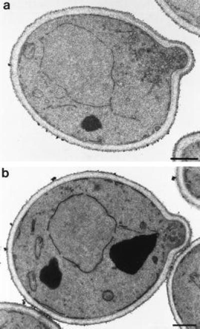
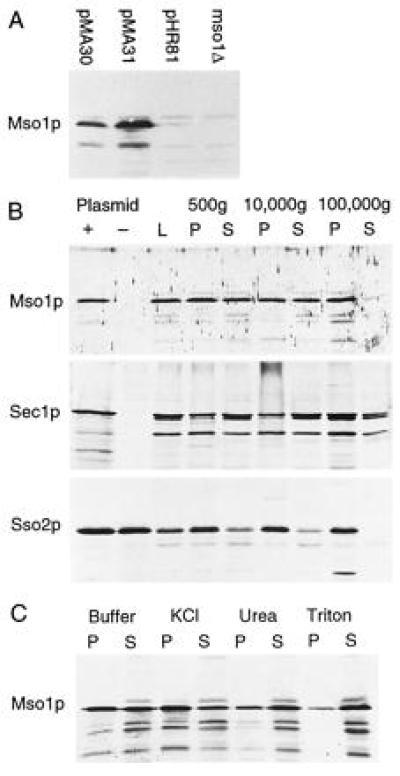
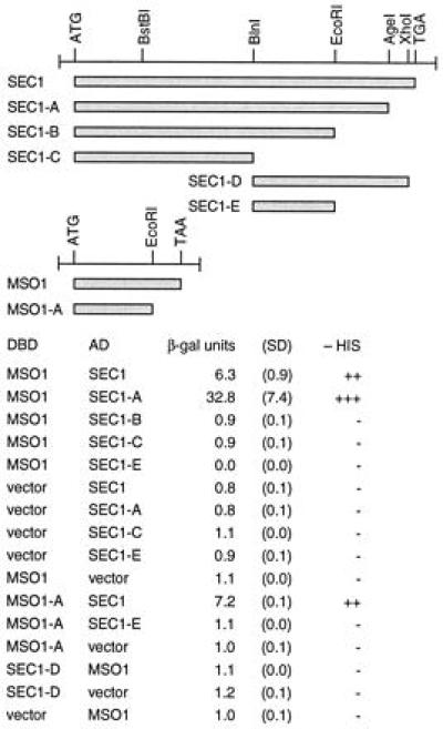
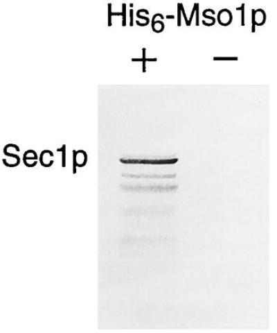
The yeast Sec1p protein functions in the docking of secretory transport vesicles to the plasma membrane. We previously have cloned two yeast genes encoding syntaxins, SSO1 and SSO2, as suppressors of the temperature-sensitive sec1-1 mutation. We now describe a third suppressor of sec1-1, which we call MSO1. Unlike SSO1 and SSO2, MSO1 is specific for sec1 and does not suppress mutations in any other SEC genes. MSO1 encodes a small hydrophilic protein that is enriched in a microsomal membrane fraction. Cells that lack MSO1 are viable, but they accumulate secretory vesicles in the bud, indicating that the terminal step in secretion is partially impaired. Moreover, loss of MSO1 shows synthetic lethality with mutations in SEC1, SEC2, and SEC4, and other synthetic phenotypes with mutations in several other late-acting SEC genes. We further found that Mso1p interacts with Sec1p both in vitro and in the two-hybrid system. These findings suggest that Mso1p is a component of the secretory vesicle docking complex whose function is closely associated with that of Sec1p.


Figures

Figure 1
Figure 1
Restriction map of MSO1. ORFs are shown as arrows. Also shown is the mso1-δ1 deletion used in one-step gene disruptions, and the insert of the smallest plasmid with suppressing activity, pMA31. The insert of pMA30 extends beyond the right end of the map.
Figure 2
Figure 2
Electron micrograph of mso1 disrupted H613 cells (a) and congenic wild-type NY179 cells (b). (Bars represent 500 nm.)
Figure 3
Figure 3
Western blots. (A) Detection of Mso1 protein in wild-type cells carrying either pMA30, pMA31, or the cloning vector pHR81. An mso1-disrupted strain is shown to the right. (B) Fractionation of yeast cell lysates by successive centrifugations at the indicated speeds. L, spheroblast lysate; P, pellet; S, supernatant. For Mso1p and Sec1p, whole cell lysates prepared in the presence and in the absence of the respective overexpression plasmid is shown (Left). There is no difference for Sso2p, because we did not need overexpression to detect this protein. (C) Membrane association of Mso1p in the 100,000 × g pellet. The pellet was treated either with Hepes buffer alone, or with buffer containing 1 M KCl, 2.5 M urea, or 1% Triton-X 100, respectively, and then separated by centrifugation at 100,000 × g.
Figure 4
Figure 4
Interaction of Mso1p and Sec1p in the two-hybrid system. Constructions that were tested are shown at the top. Results are shown as β-galactosidase units, with SD of three independent transformants in parentheses, and as growth in the absence of histidine. AD, activating domain; DBD, DNA binding domain.
Figure 5
Figure 5
In vitro association of Sec1p and Mso1p. A lysate from the Sec1p-overproducing strain was incubated with Ni-NTA-agarose beads in the presence or in the absence of His6-Mso1p. Bound protein was analyzed in a Western blot using an antiserum against Sec1p.

References

    1. Jamieson J, Palade G. J Cell Biol. 1968;39:589–603. - PMC - PubMed
    1. Palade G. Science. 1975;189:347–358. - PubMed
    1. Novick P J, Schekman R. Proc Natl Acad Sci USA. 1979;76:1858–1862. - PMC - PubMed
    1. Novick P J, Ferro S, Schekman R. Cell. 1981;25:461–469. - PubMed
    1. Aalto M K, Ruohonen L, Hosono K, Keränen S. Yeast. 1991;7:643–650. , and corrigendum (1992) 8, 587–588. - PubMed

Publication types

MeSH terms