

Induction of host signal transduction pathways by Helicobacter pylori
- PMID: 9207137
- PMCID: PMC23867
- DOI: 10.1073/pnas.94.14.7595
Induction of host signal transduction pathways by Helicobacter pylori
Abstract
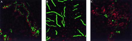
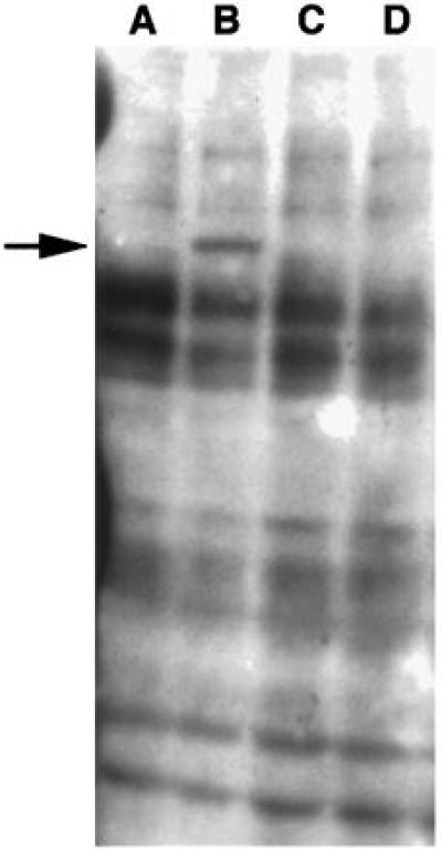
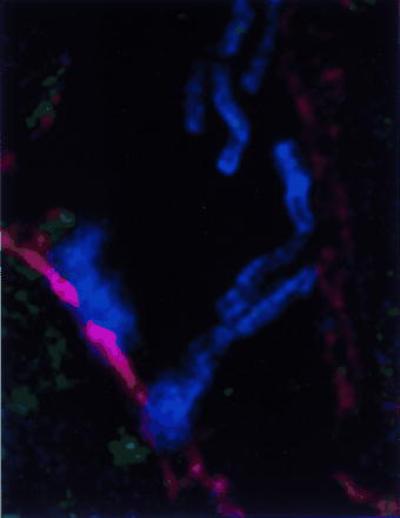
Adherence of Helicobacter pylori to cultured gastric epithelial cells is associated with several cellular events, including the tyrosine phosphorylation of a 145-kDa host protein; the reorganization of the host cell actin and associated cellular proteins, like vasodilator-stimulated phosphoprotein, adjacent to the attached bacterial cell; and the subsequent release of the cytokine, interleukin 8 (IL-8). H. pylori isolated from patients with ulcer disease and gastric cancer contain a DNA insertion, the cag pathogenicity island (PAI), that is not present in bacteria isolated from individuals with asymptomatic infection. Mutations in a number of PAI genes abolish tyrosine phosphorylation and IL-8 synthesis but not the cytoskeletal rearrangements. Kinase inhibition studies suggest there are two distinct pathways operative in stimulating IL-8 release from host cells and one of these H. pylori pathways is independent of the tyrosine phosphorylation step.
Figures
References
-
- Logan R P. Lancet. 1994;344:1078–1079. - PubMed
-
- McColm, A. A., Bagshaw, J., O’Malley, C. & McLaren, A. (1991) Microb. Ecol. Health Dis. 4, Suppl., S145 (abstr.).
-
- Weel J F, van der Hulst R W, Gerrits Y, Roorda P, Feller M, Dankert J, Tytgat G N, van der Ende A. J Infect Dis. 1996;173:1171–1175. - PubMed
Publication types
MeSH terms
Substances
Grants and funding
LinkOut - more resources
Full Text Sources
Molecular Biology Databases
