. 1997 Jul 8;94(14):7633-8.
doi: 10.1073/pnas.94.14.7633.

Transmembrane topology of a CLC chloride channel

Affiliations

Transmembrane topology of a CLC chloride channel

T Schmidt-Rose et al. Proc Natl Acad Sci U S A. .

Abstract

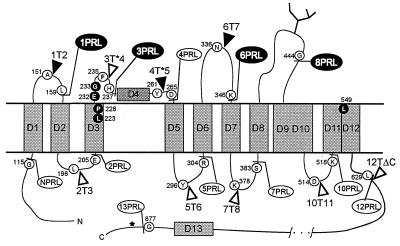
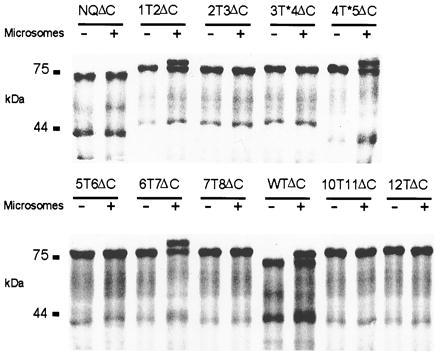
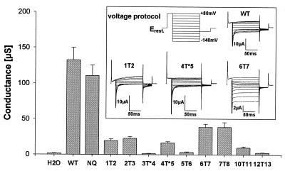
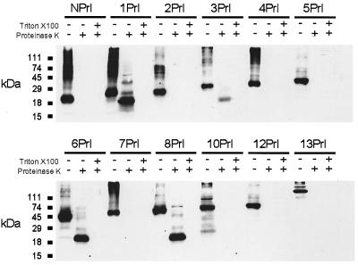
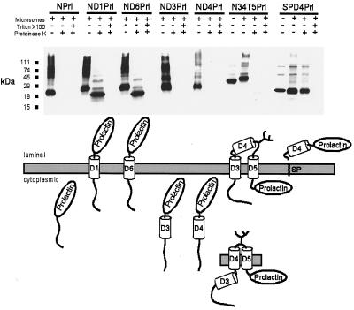
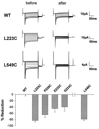
CLC chloride channels form a large and conserved gene family unrelated to other channel proteins. Knowledge of the transmembrane topology of these channels is important for understanding the effects of mutations found in human myotonia and inherited hypercalciuric kidney stone diseases and for the interpretation of structure-function studies. We now systematically study the topology of human ClC-1, a prototype CLC channel that is defective in human myotonia. Using a combination of in vitro glycosylation scanning and protease protection assays, we show that both N and C termini face the cytoplasm and demonstrate the presence of 10 (or less likely 12) transmembrane spans. Difficult regions were additionally tested by inserting cysteines and probing the effect of cysteine-modifying reagents on ClC-1 currents. The results show that D3 crosses the membrane and D4 does not, and that L549 between D11 and D12 is accessible from the outside. Further, since the modification of cysteines introduced between D11 and D12 and at the extracellular end of D3 strongly affect ClC-1 currents, these regions are suggested to be important for ion permeation.


Figures

Figure 1
Figure 1
Topological model of human ClC-1 illustrating the constructs used in this work. Hydrophobic domains are labeled D1 to D13 according to the original nomenclature for ClC-0 (5) based on Kyte–Doolittle hydropathy analysis. For amino acid sequence, see ref. . WT-glycosylation is represented by a branched line in loop D8/D9. An unused glycosylation site after D13 is indicated by an asterisk. Insertion sites of glycosylation transplants are indicated by triangles (solid = glycosylation positive), fusion sites with prolactin by ball-and-stick (solid = proteinase protection positive) after the amino acids shown in open circles. Amino acids in black have been replaced by cysteines and are sensitive to cysteine specific reagents.
Figure 2
Figure 2
Autoradiograph of in vitro translation products with glycosylation sites engineered between hydrophobic domains. RNA was translated in reticulocyte lysate in absence (−) or presence (+) of microsomal membranes. All constructs are shortened at the C terminus as indicated by ΔC. WTΔC contains the native glycosylation site. In NQΔC the glycosylation site is removed by mutating N430 to Q. “T” in other constructs indicates the standard glycosylation transplant, “T*” the longer variant, and the flanking numbers the respective hydrophobic domains. Numbers at left indicate the molecular mass in kDa.
Figure 3
Figure 3
Two-electrode voltage clamp experiments of Xenopus oocytes expressing full-length glycosylation constructs. Mean conductance at −40 mV of WT ClC-1 and its mutants in comparison to water injected control oocytes (mean ± SEM, n = 4 to 9). (Inset) Pulse protocol and typical current traces for WT and constructs 1T2, 4T*5, and 6T7.
Figure 4
Figure 4
Proteinase protection assay. Increasing portions of ClC-1 starting at the N terminus (N) were fused C-terminally to a prolactin reporter-epitope (Prl). Numbers indicate the last hydrophobic domain of ClC-1 that is included in the fusion protein. In vitro translation was performed in the presence of pancreatic microsomes. Products were treated with proteinase K with (+) or without (−) Triton X-100. Controls were treated equivalently, but proteinase K and detergent were omitted (−/−).
Figure 5
Figure 5
Testing of individual domains for their topogenic activity. Modular fusions of individual domains with the ClC-1 N terminus and the prolactin reporter epitope. In construct SPD4Prl the N terminus is formed by the signal sequence of pre-prolactin (SP). (Upper) Immunoblot of in vitro translation products treated as described above. (Lower) Schematic drawings of topologies compatible with the experimental results.
Figure 6
Figure 6
Effects of MTSES on Xenopus oocytes expressing ClC-1 WT or cysteine mutants indicated in Fig. 1. (Upper) Examples of current traces. (Lower) Relative changes in conductance at −40 mV 3 min after application of 10 mM MTSES (mean ± SEM, n = 5 to 9) and subsequent washout.

References

    1. Fujita N, Mori H, Yura T, Ishihama A. Nucleic Acids Res. 1994;22:1637–1639. - PMC - PubMed
    1. Greene J R, Brown N H, DiDomenico B J, Kaplan J, Eide D J. Mol Gen Genet. 1993;241:542–553. - PubMed
    1. Hechenberger M, Schwappach B, Fischer W N, Frommer W B, Jentsch T J, Steinmeyer K. J Biol Chem. 1996;271:33632–33638. - PubMed
    1. Steinmeyer K, Ortland C, Jentsch T J. Nature (London) 1991;354:301–304. - PubMed
    1. Jentsch T J, Steinmeyer K, Schwarz G. Nature (London) 1990;348:510–514. - PubMed

Publication types

LinkOut - more resources