. 1997 Jul 14;138(1):17-36.
doi: 10.1083/jcb.138.1.17.

A small conserved domain in the yeast Spa2p is necessary and sufficient for its polarized localization

Affiliations

A small conserved domain in the yeast Spa2p is necessary and sufficient for its polarized localization

R A Arkowitz et al. J Cell Biol. .

Abstract

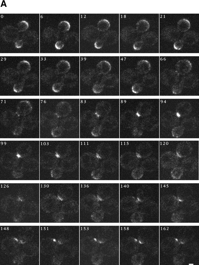
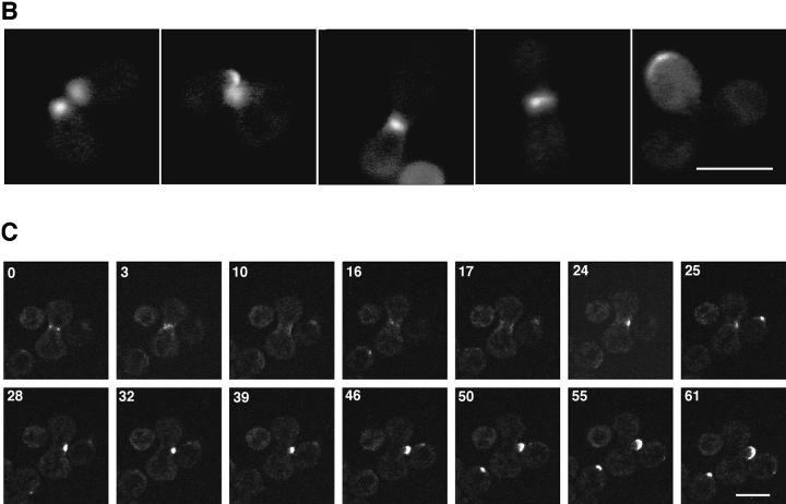
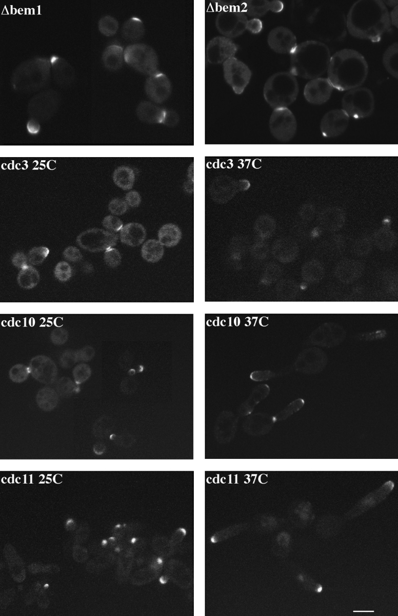
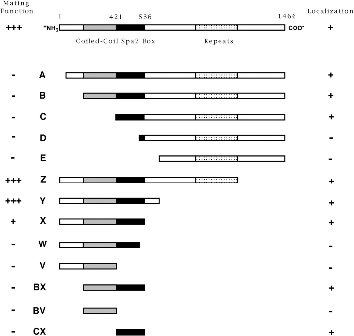
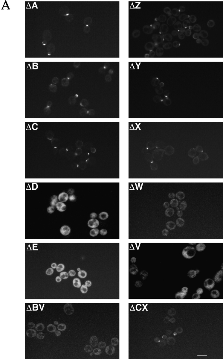
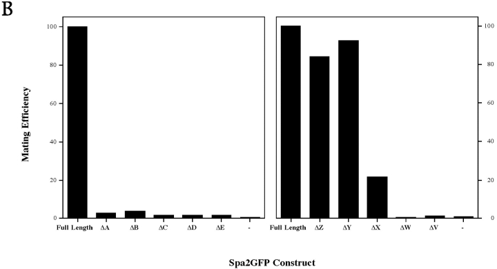
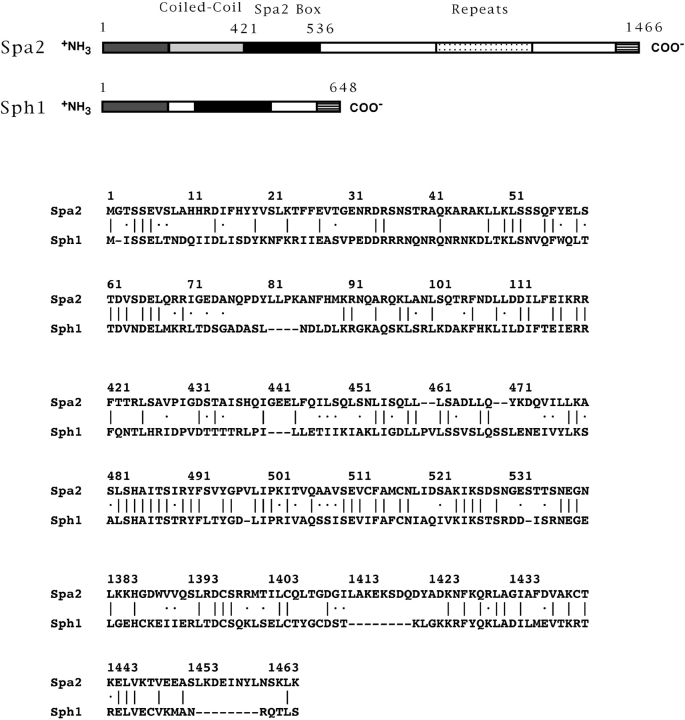
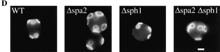
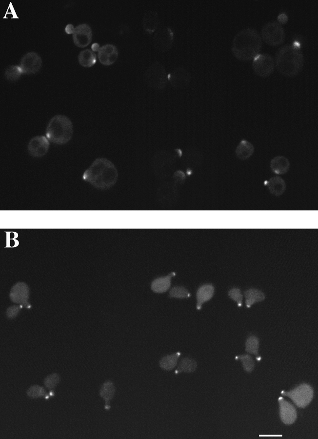
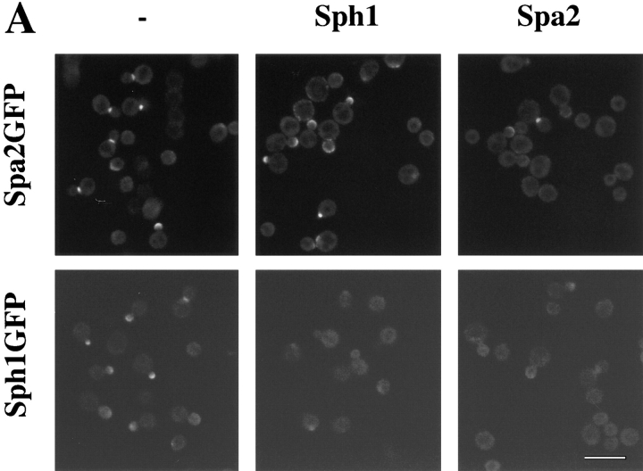
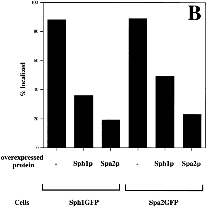
SPA2 encodes a yeast protein that is one of the first proteins to localize to sites of polarized growth, such as the shmoo tip and the incipient bud. The dynamics and requirements for Spa2p localization in living cells are examined using Spa2p green fluorescent protein fusions. Spa2p localizes to one edge of unbudded cells and subsequently is observable in the bud tip. Finally, during cytokinesis Spa2p is present as a ring at the mother-daughter bud neck. The bud emergence mutants bem1 and bem2 and mutants defective in the septins do not affect Spa2p localization to the bud tip. Strikingly, a small domain of Spa2p comprised of 150 amino acids is necessary and sufficient for localization to sites of polarized growth. This localization domain and the amino terminus of Spa2p are essential for its function in mating. Searching the yeast genome database revealed a previously uncharacterized protein which we name, Sph1p (a2p omolog), with significant homology to the localization domain and amino terminus of Spa2p. This protein also localizes to sites of polarized growth in budding and mating cells. SPH1, which is similar to SPA2, is required for bipolar budding and plays a role in shmoo formation. Overexpression of either Spa2p or Sph1p can block the localization of either protein fused to green fluorescent protein, suggesting that both Spa2p and Sph1p bind to and are localized by the same component. The identification of a 150-amino acid domain necessary and sufficient for localization of Spa2p to sites of polarized growth and the existence of this domain in another yeast protein Sph1p suggest that the early localization of these proteins may be mediated by a receptor that recognizes this small domain.


Figures

Figure 1
Figure 1
(A) Complementation of spa2 mating defect by SPA2GFP. Δspa2 mutants with SPA2 (RAY786), SPA2GFP (RAY651), or an empty plasmid (RAY698) integrated at the URA3 locus were mated with the enfeebled mating tester strain (JY426). Diploids were selected on −ura, −lys plates. Mating efficiency with SPA2 (30.9%) was set to 100% efficiency. (B) Complementation of spa2 bipolar budding defect by SPA2GFP. Wild-type diploids (SEY6210/11) and homozygous Δspa2 diploids with either SPA2GFP (RAY775) or an empty plasmid (RAY776) integrated at the URA3 locus growing exponentially were stained with Calcofluor as described in Materials and Methods, and budding pattern was analyzed. For each strain, the position of the bud relative to the birth scar (see inset) was scored for ∼100 cells with two or more bud scars. (C) Localization of Spa2GFP at sites of polarized growth. Confocal microscopy of living cells with SPA2GFP integrated at URA3 (RAY416) at different stages in cell cycle. Note unbudded cells with patch of Spa2GFP, small- and medium-sized buds with Spa2GFP fluorescence on the periphery of the tip, and a cell undergoing cytokinesis with a “bar” of Spa2GFP at the mother–daughter bud neck. Bar, 5 μm.
Figure 2
Figure 2
(A) Dynamics of Spa2GFP localization in living cells. Spa2GFP distribution in two cells (RAY416) at 6-min intervals. The Spa2GFP fluorescence in two cells is in different focal planes and the upper cell is in focus throughout the time course. Note the appearance at 76 min of a ring at the mother bud neck. Cells divided during and after confocal time course. (B) Localization of Spa2GFP in mating cells. Confocal microscopy of five representative mating pairs (RAY416 and RAY674) at different stages of mating/zygote formation. The diffuse background is due to the high gain setting to allow visualization of the entire cell. (C) Movement of Spa2GFP in budding zygotes. Time course of zygotes from RAY416 mated to RAY674. In the center of each panel is a dumbbell-shaped zygote with a separate round cell to its right and left. Note the ring of Spa2GFP fluorescence at the a/α shmoo neck at 0 min. Bars: (A and B) 1 μm; (C) 5 μm.
Figure 2
Figure 2
(A) Dynamics of Spa2GFP localization in living cells. Spa2GFP distribution in two cells (RAY416) at 6-min intervals. The Spa2GFP fluorescence in two cells is in different focal planes and the upper cell is in focus throughout the time course. Note the appearance at 76 min of a ring at the mother bud neck. Cells divided during and after confocal time course. (B) Localization of Spa2GFP in mating cells. Confocal microscopy of five representative mating pairs (RAY416 and RAY674) at different stages of mating/zygote formation. The diffuse background is due to the high gain setting to allow visualization of the entire cell. (C) Movement of Spa2GFP in budding zygotes. Time course of zygotes from RAY416 mated to RAY674. In the center of each panel is a dumbbell-shaped zygote with a separate round cell to its right and left. Note the ring of Spa2GFP fluorescence at the a/α shmoo neck at 0 min. Bars: (A and B) 1 μm; (C) 5 μm.
Figure 3
Figure 3
Spa2GFP localization in bem and septin mutants. Δbem1 (RAY712), Δbem2 (RAY836), cdc3-1 (RAY696), cdc10-1 (RAY685), and cdc11-1 (RAY691) strains with SPA2GFP integrated at the URA3 locus were grown at 25°C. Exponentially growing cultures of the septin mutants were shifted to 37°C for 3 h, and cells were examined by confocal microscopy. Note large cells in the bem mutants and the Spa2GFP fluorescence at both the bud tip and mother–daughter bud neck. Bar, 5 μm.
Figure 4
Figure 4
Diagram of Spa2GFP deletion constructs. Deletions ΔA, ΔB, ΔC, ΔD, and ΔE contain Spa2p residues 88, 288, 397, 511, and 625, respectively, to residue 1,463. Deletions ΔZ, ΔY, ΔX, ΔW, and ΔV contain Spa2p residue 1 to residue 1,074, 655, 549, 511, and 396, respectively. Constructs ΔBX, ΔBV, and ΔCX contain Spa2p residues 288–549, 288–396, and 397–549, respectively. All constructs contain Spa2p fused to GFP and were integrated at the URA3 locus in either SEY6210 for localization studies or JC-J9 for mating function analysis. See Fig. 5 A for localization data and Fig. 5 B for results from mating assays.
Figure 5
Figure 5
(A) Localization of Spa2GFP deletion mutants. Exponentially growing cultures of SEY6210 with deletion constructs integrated at the URA3 locus were examined by confocal microscopy. In ΔA, ΔB, ΔC, ΔZ, ΔY, ΔX, and ΔCX, note both bud tip and mother–daughter bud neck localization of Spa2GFP deletions. Panels ΔD, ΔE, ΔC, ΔW, ΔV, and ΔCX show general Spa2GFP fluorescence. (B) Mating function of Spa2GFP deletion mutants. The spa2 mutant (JC-J9) with SPA2GFP deletion constructs integrated at the URA3 locus was mated with an enfeebled mating partner (JY429), and diploids were selected on −met, −ura plates. In each experiment, mating of JC-J9 carrying full-length Spa2GFP was taken as 100% efficiency (absolute efficiency of 11.2% [left] and 7.9% [right]). Bar, 5 μm.
Figure 5
Figure 5
(A) Localization of Spa2GFP deletion mutants. Exponentially growing cultures of SEY6210 with deletion constructs integrated at the URA3 locus were examined by confocal microscopy. In ΔA, ΔB, ΔC, ΔZ, ΔY, ΔX, and ΔCX, note both bud tip and mother–daughter bud neck localization of Spa2GFP deletions. Panels ΔD, ΔE, ΔC, ΔW, ΔV, and ΔCX show general Spa2GFP fluorescence. (B) Mating function of Spa2GFP deletion mutants. The spa2 mutant (JC-J9) with SPA2GFP deletion constructs integrated at the URA3 locus was mated with an enfeebled mating partner (JY429), and diploids were selected on −met, −ura plates. In each experiment, mating of JC-J9 carrying full-length Spa2GFP was taken as 100% efficiency (absolute efficiency of 11.2% [left] and 7.9% [right]). Bar, 5 μm.
Figure 6
Figure 6
Alignment of Spa2p and Sph1p sequences. A schematic representation of regions of identity as identified by the BLAST algorithm (Altschul et al., 1990) is shown. Aligned sequences are shown below. Sph1p is 15% identical to Spa2p over the entire protein. ∣, identical amino acids; ·, similar amino acids. A, B, and C, three regions of similarity between Spa2p and Sph1p. Numbers correspond to amino acid residue numbers of Spa2p and Sph1p.
Figure 7
Figure 7
(A) Deletion of sph1 has no effect on mating efficiency. Exponential cultures of WT (RAY697), Δspa2 (RAY698), Δsph1 (RAY709), and Δspa2 Δsph1 (RAY711) were mated with an enfeebled mating tester strain (JY426) as described in Materials and Methods, and diploids were selected on −lys, −ura plates. Wild-type (WT) mating efficiency (4.4%) was set to 100% efficiency. (B) SPH1 can partially complement for mating deficiency of Δspa2 (RAY574). WT (RAY697), Δspa2 with SPH1GFP (RAY703), and Δspa2 (RAY698) were mated with an enfeebled mating tester strain (JY426) as described in Materials and Methods, and diploids were selected on −lys, −ura plates. WT mating efficiency (6.5%) was normalized to 100% efficiency. (C) SPH1 is required for bipolar bud site selection. Wild-type (SEY6210/11), homozygous Δspa2 (RAY616), homozygous Δsph1 (RAY618), and homozygous Δspa2 Δsph1 (RAY620) exponentially growing diploids were stained with Calcofluor as described in Materials and Methods, and budding pattern was analyzed. For each strain, the position of the bud relative to the birth scar (see inset) was scored for ∼150 cells with two or more bud scars. (D) Budding pattern of homozygous diploids sph1 and spa2 mutants. Representative fluorescence microscopy pictures of cells quantitated in C. Bar, 2.5 μm.
Figure 7
Figure 7
(A) Deletion of sph1 has no effect on mating efficiency. Exponential cultures of WT (RAY697), Δspa2 (RAY698), Δsph1 (RAY709), and Δspa2 Δsph1 (RAY711) were mated with an enfeebled mating tester strain (JY426) as described in Materials and Methods, and diploids were selected on −lys, −ura plates. Wild-type (WT) mating efficiency (4.4%) was set to 100% efficiency. (B) SPH1 can partially complement for mating deficiency of Δspa2 (RAY574). WT (RAY697), Δspa2 with SPH1GFP (RAY703), and Δspa2 (RAY698) were mated with an enfeebled mating tester strain (JY426) as described in Materials and Methods, and diploids were selected on −lys, −ura plates. WT mating efficiency (6.5%) was normalized to 100% efficiency. (C) SPH1 is required for bipolar bud site selection. Wild-type (SEY6210/11), homozygous Δspa2 (RAY616), homozygous Δsph1 (RAY618), and homozygous Δspa2 Δsph1 (RAY620) exponentially growing diploids were stained with Calcofluor as described in Materials and Methods, and budding pattern was analyzed. For each strain, the position of the bud relative to the birth scar (see inset) was scored for ∼150 cells with two or more bud scars. (D) Budding pattern of homozygous diploids sph1 and spa2 mutants. Representative fluorescence microscopy pictures of cells quantitated in C. Bar, 2.5 μm.
Figure 8
Figure 8
(A) Sph1p localizes to sites of polarized growth in budding cells. Exponentially grown cells with SPH1GFP integrated at URA3 (RAY699) were analyzed by confocal microscopy. Immunoblot analyses of RAY699 revealed that a GFP fusion protein of the correct molecular mass (∼100 kD) was expressed. (B) Sph1p localizes to sites of polarized growth in shmoos. Exponentially grown Mata Δsph1 cells with SPH1GFP integrated at URA3 (RAY875) were treated with α-factor, as described in Materials and Methods, fixed, and viewed by confocal microscopy. Note that background fluorescence in cells is due to Ade fluorophore from ade2 mutation. Bar, 5 μm.
Figure 9
Figure 9
Localization of Spa2GFP or Sph1GFP is blocked by overexpression of either protein. A strain with SPA2GFP integrated at the URA3 locus (RAY416) was transformed with either pRS425 (), pRS425TPISPH1myc (Sph1), or pRS425TPISPA2myc (Spa2). A strain with SPH1GFP integrated at the URA3 locus (RAY699) was transformed with either pRS425 (), pRS425TPISPH1myc (Sph1), or pRS425TPISPA2myc (Spa2). Exponentially grown cultures were analyzed by confocal microscopy for GFP fluorescence. Each panel shows a representative field of view. (B) Quantitation of cells with localized Spa2GFP or Sph1GFP. For each condition, 300 cells were scored for the presence of fluorescence localized to regions of polarized growth using either fluorescence or confocal microscopy. Bar, 10 μm.
Figure 9
Figure 9
Localization of Spa2GFP or Sph1GFP is blocked by overexpression of either protein. A strain with SPA2GFP integrated at the URA3 locus (RAY416) was transformed with either pRS425 (), pRS425TPISPH1myc (Sph1), or pRS425TPISPA2myc (Spa2). A strain with SPH1GFP integrated at the URA3 locus (RAY699) was transformed with either pRS425 (), pRS425TPISPH1myc (Sph1), or pRS425TPISPA2myc (Spa2). Exponentially grown cultures were analyzed by confocal microscopy for GFP fluorescence. Each panel shows a representative field of view. (B) Quantitation of cells with localized Spa2GFP or Sph1GFP. For each condition, 300 cells were scored for the presence of fluorescence localized to regions of polarized growth using either fluorescence or confocal microscopy. Bar, 10 μm.

References

    1. Adams AE, Pringle JR. Relationship of actin and tubulin distribution to bud growth in wild-type and morphogenetic-mutant Saccharomyces cerevisiae. . J Cell Biol. 1984;98:934–945. - PMC - PubMed
    1. Altschul SF, Gish W, Miller W, Myers EW, Lipman DJ. Basic local alignment search tool. J Mol Biol. 1990;215:403–410. - PubMed
    1. Amatruda JF, Cooper JA. Purification, characterization, and immunofluorescence localization of Saccharomyces cerevisiaecapping protein. J Cell Biol. 1992;117:1067–1076. - PMC - PubMed
    1. Anderson K. One signal, two body axes. Science (Wash DC) 1995;269:489–490. - PubMed
    1. Baudin A, Ozier-Kalogeropoulos O, Denouel A, Lacroute R, Cullin C. A simple and efficient method for direct gene deletion in Saccharomyces cerevisiae. . Nucleic Acids Res. 1993;21:3329–3330. - PMC - PubMed

MeSH terms

Associated data