. 1997 Jul 14;138(1):65-80.
doi: 10.1083/jcb.138.1.65.

A novel class of RanGTP binding proteins

Affiliations

A novel class of RanGTP binding proteins

D Görlich et al. J Cell Biol. .

Abstract

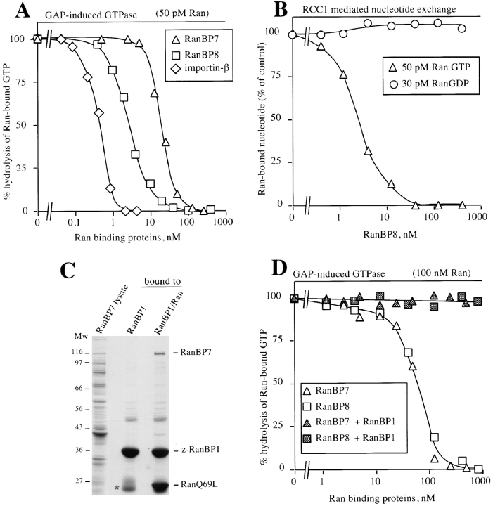
The importin-alpha/beta complex and the GTPase Ran mediate nuclear import of proteins with a classical nuclear localization signal. Although Ran has been implicated also in a variety of other processes, such as cell cycle progression, a direct function of Ran has so far only been demonstrated for importin-mediated nuclear import. We have now identified an entire class of approximately 20 potential Ran targets that share a sequence motif related to the Ran-binding site of importin-beta. We have confirmed specific RanGTP binding for some of them, namely for two novel factors, RanBP7 and RanBP8, for CAS, Pse1p, and Msn5p, and for the cell cycle regulator Cse1p from Saccharomyces cerevisiae. We have studied RanBP7 in more detail. Similar to importin-beta, it prevents the activation of Ran's GTPase by RanGAP1 and inhibits nucleotide exchange on RanGTP. RanBP7 binds directly to nuclear pore complexes where it competes for binding sites with importin-beta, transportin, and apparently also with the mediators of mRNA and U snRNA export. Furthermore, we provide evidence for a Ran-dependent transport cycle of RanBP7 and demonstrate that RanBP7 can cross the nuclear envelope rapidly and in both directions. On the basis of these results, we propose that RanBP7 might represent a nuclear transport factor that carries an as yet unknown cargo, which could apply as well for this entire class of related RanGTP-binding proteins.


Figures

Figure 1
Figure 1
A 120-kD Ran-binding protein copurifies with importin-β. (A) A Xenopus egg extract (high speed supernatant, lane 1) was subjected to binding to an immobilized IBB domain at 200 mM NaCl. The starting material (lane 1) and the bound fractions (lanes 2–4) were analyzed by SDS-PAGE followed by Coomassie staining. Two bands are specifically recovered in the IBB-bound fraction, importin-β, and a copurifying 120-kD protein that is referred to here as RanBP7. Note, the addition of 10 μM RanQ69L GTP or nonimmobilized IBB competitor (50 μM) prevent binding of both importin-β and RanBP7 (lanes 3 and 4, respectively). (B) RanBP7 was expressed in E. coli. A lysate was prepared and subjected to binding to an IBB column or to an IBB column to which importin-β had been prebound. The starting material and the bound fractions were analyzed by SDS-PAGE followed by Coomassie staining. Note, binding of RanBP7 to the IBB domain is via importin-β. (C) A digitonin (50 μg/ml) extract from HeLa cells was prepared and bound to an immobilized IBB domain as in A. Analysis was by Coomassie staining and by immunoblotting with affinity purified antibodies raised against RanBP7, importin-β, importin-α (Rch1p), and Ran. Again, two bands were recovered specifically in the bound fraction: importin-β and a 120-kD protein that cross-reacts with the anti–Xenopus RanBP7 antibody. (D) A blot of HeLa cell extract and the corresponding IBB-bound fraction (as in C) was probed with a RanBP1/Ran γ-[32P]GTP complex, followed by autoradiography. In the IBB-bound fraction, both importin-β and RanBP7 give a strong signal. In the crude lysate, importin-β and a double band of 120 kD is detected. The latter consists probably of several Ran-binding proteins, in addition to RanBP7.
Figure 2
Figure 2
The RanBP7–importin-β interaction is regulated by Ran and appears unrelated to NLS-mediated nuclear protein import. (A) A Xenopus egg extract (high speed supernatant) was bound either to an immobilized BSA–NLS conjugate or an IBB domain. The starting material and the bound fractions were analyzed by immunoblots against RanBP7, importin-β, and -α. Note, whereas equal amounts of importin-β were found in both bound fractions, RanBP7 was recovered only with IBB column but hardly at all with the NLS conjugate. (B) z-Tagged RanBP7 was prebound to IgG Sepharose and used to bind importin-β out of a Xenopus egg extract (z is an IgG binding domain from Straphylococcus aureus protein A). Binding was with or without the addition of 10 μM RanQ69L GTP. Starting material and bound fractions were analyzed by immunoblotting against Ran and importin-β, but the z-tag is also detected by this procedure. Note that importin-β but no Ran bound to RanBP7 in the absence of RanQ69L. If however, the Ran mutant was added, importin-β binding was abolished, and instead, Ran (Q69L) was recovered in the bound fraction. (C) RanBP7 was expressed in E. coli. A lysate was prepared and subjected to binding to z-tagged importin-β that had been preabsorbed to IgG Sepharose. Elution from the beads was either with SDS, which also releases z–importin-β from the column, or with RanQ69L GTP which dissociates RanBP7 from importin-β. *, dimerized Ran; **, IgG light chain.
Figure 2
Figure 2
The RanBP7–importin-β interaction is regulated by Ran and appears unrelated to NLS-mediated nuclear protein import. (A) A Xenopus egg extract (high speed supernatant) was bound either to an immobilized BSA–NLS conjugate or an IBB domain. The starting material and the bound fractions were analyzed by immunoblots against RanBP7, importin-β, and -α. Note, whereas equal amounts of importin-β were found in both bound fractions, RanBP7 was recovered only with IBB column but hardly at all with the NLS conjugate. (B) z-Tagged RanBP7 was prebound to IgG Sepharose and used to bind importin-β out of a Xenopus egg extract (z is an IgG binding domain from Straphylococcus aureus protein A). Binding was with or without the addition of 10 μM RanQ69L GTP. Starting material and bound fractions were analyzed by immunoblotting against Ran and importin-β, but the z-tag is also detected by this procedure. Note that importin-β but no Ran bound to RanBP7 in the absence of RanQ69L. If however, the Ran mutant was added, importin-β binding was abolished, and instead, Ran (Q69L) was recovered in the bound fraction. (C) RanBP7 was expressed in E. coli. A lysate was prepared and subjected to binding to z-tagged importin-β that had been preabsorbed to IgG Sepharose. Elution from the beads was either with SDS, which also releases z–importin-β from the column, or with RanQ69L GTP which dissociates RanBP7 from importin-β. *, dimerized Ran; **, IgG light chain.
Figure 3
Figure 3
Molecular cloning of Xenopus RanBP7 and human RanBP8 and their similarities to S. cerevisiae Nmd5p. Xenopus RanBP7 was cloned from an oocyte library using partial peptide sequence information from the purified protein. RanBP8 was found in the data base as an expressed human sequence tag similar to RanBP7 and was subsequently cloned from HeLa cDNA. Shown are the aligned amino acid sequences from Xenopus RanBP7, human RanBP8, and S. cerevisiae Nmd5p. Residues identical in all three proteins are indicated by #, and similar amino acids by *. The sequence data for Xenopus RanBP7 and human RanBP8 are available from GenBank/EMBL/DDBJ under accession numbers U71082 and U77494, respectively.
Figure 4
Figure 4
The RanBP7/importin-β/Cse1p superfamily. (A) Multiple alignment of amino acids 6–141 from RanBP7 with the NH2 termini of other proteins that have a similar NH2-terminal sequence motif. Positions in red match the consensus. The consensus was defined by the two most frequent amino acids in each position. Proteins named in blue have been shown to bind Ran (see main text and Fig. 5). S. pombe Crm1p and HRC1004 also match the motif but are not shown in the figure. (For accession numbers see Materials and Methods.) (B) Relationship between the proteins shown in A. The tree was calculated from the entire coding sequences and not just from the NH2 termini. Proteins named in red have been shown to bind RanGTP (see also below).
Figure 4
Figure 4
The RanBP7/importin-β/Cse1p superfamily. (A) Multiple alignment of amino acids 6–141 from RanBP7 with the NH2 termini of other proteins that have a similar NH2-terminal sequence motif. Positions in red match the consensus. The consensus was defined by the two most frequent amino acids in each position. Proteins named in blue have been shown to bind Ran (see main text and Fig. 5). S. pombe Crm1p and HRC1004 also match the motif but are not shown in the figure. (For accession numbers see Materials and Methods.) (B) Relationship between the proteins shown in A. The tree was calculated from the entire coding sequences and not just from the NH2 termini. Proteins named in red have been shown to bind RanGTP (see also below).
Figure 5
Figure 5
Human CAS and the yeast proteins Pse1p, Cse1p, and Msn5p are Ran (Gsp1p) binding. Indicated proteins were expressed in E. coli, and the total lysates were separated by SDS-PAGE and transferred onto nitrocellulose. The blot in A was probed with 10 pM Ran γ-[32P]GTP in the presence of a 5,000-fold molar excess of RanBP1, followed by autoradiography. The negative control was a lysate from E. coli expressing importin-α. The blots in B were probed with 10 pM Gsp1p γ-[32P]GTP (Gsp1p is Ran from S. cerevisiae). The negative control was importin-α, as in A. Pse1p 1–257 and Pse1p 1–409 are fragments of Pse1p comprising of the NH2-terminal 257 or 409 residues. Msn5p was expressed as an NH2-terminal fragment comprised of the NH2-terminal 682 residues.
Figure 6
Figure 6
Enzymatic interactions of RanBP7 and RanBP8 with Ran. (A) RanBP7 and RanBP8 inhibit GAP induction of GTPase activity of Ran. 50 pM Ran γ-[32P]GTP was preincubated for 30 min with the indicated concentrations of importin-β, RanBP7, and RanBP8. 5 nM Rna1p (RanGAP from S. pombe) was added, and GTP hydrolysis was allowed for 5 min. Hydrolysis of Ran-bound GTP was measured as released γ-[32P]phosphate. When using a very low Ran concentration as in this experiment, the method can be used to estimate the K D of the Ran-binding proteins for RanGTP. These are ∼0.8, 25, and 3 nM for importin-β, RanBP7, and RanBP8, respectively. (B) Effect of RanBP8 on guanine nucleotide exchange on Ran. 30 pM Ran α-[32P]GDP or 50 pM Ran α-[32P]GTP were preincubated for 30 min with indicated concentrations of RanBP8. The exchange reaction was started by addition of 0.2 mM unlabeled GDP plus 2 nM of the exchange factor RCC1. After 5 min, Ran-bound 32P-labeled nucleotide was measured in a filter binding assay. Note, RanBP8 inhibits nucleotide exchange on RanGTP but not on RanGDP. (C) RanBP7 can form a trimeric RanBP7/RanGTP/RanBP1 complex. z-Tagged RanBP1 was prebound to IgG Sepharose. A RanBP7 lysate was subjected to binding to either immobilized RanBP1 alone or to an immobilized RanBP1/RanGTP complex. Elution was with SDS, which also releases the z-RanBP1 from the IgG Sepharose. Note, RanBP7 does not bind to RanBP1 alone, but it is specifically recovered with the RanBP1/RanGTP complex. *, IgG light chain that leaked from the column. (D) The GAP resistance of the RanBP7/RanGTP and the RanBP8/RanGTP complex is relieved by RanBP1. 100 nM RanGTP was preincubated for 30 min with indicated concentrations of RanBP7 or RanBP8. Buffer or 200 nM RanBP1 were added, followed immediately by 200 nM Rna1p. GTP hydrolysis was allowed for 5 min and measured as in A.
Figure 7
Figure 7
Characterization of RanBP7 transport between nucleus and cytoplasm. (A) RanBP7 was translated in the reticulocyte system in the presence of [35S]methionine and mixed with [14C]BSA, which served as an injection control. The mixture was then injected into Xenopus laevis oocyte nuclei either alone or together with 40 μM Rna1p, or with Rna1p and 80 μM RanQ69L. Proteins were extracted 5 or 90 min after injection. T, C, and N, indicate proteins extracted from total oocytes or after dissection from cytoplasmic or nuclear fractions, respectively. Proteins were analyzed by SDS-PAGE followed by fluorography. (B) Nuclei of Xenopus oocytes were injected either with buffer (control), or 10 μM Rna1p. Oocytes were dissected 6 h later, and the distribution of endogenous proteins was analyzed by SDS-PAGE followed by Coomassie staining, or by Western blotting with RanBP7 and importin-α antibodies. In the control, RanBP7 and importin-α are predominantly cytoplasmic. Nearly 50% of RanBP7 and ∼20% of importin-α accumulated in the nucleus, after nuclear Rna1p injection that inhibits re-export of RanBP7 and importin-α. *, Hemoglobin from the injection control. Rna1p stays nuclear after nuclear injection as judged by Western blotting (not shown).
Figure 7
Figure 7
Characterization of RanBP7 transport between nucleus and cytoplasm. (A) RanBP7 was translated in the reticulocyte system in the presence of [35S]methionine and mixed with [14C]BSA, which served as an injection control. The mixture was then injected into Xenopus laevis oocyte nuclei either alone or together with 40 μM Rna1p, or with Rna1p and 80 μM RanQ69L. Proteins were extracted 5 or 90 min after injection. T, C, and N, indicate proteins extracted from total oocytes or after dissection from cytoplasmic or nuclear fractions, respectively. Proteins were analyzed by SDS-PAGE followed by fluorography. (B) Nuclei of Xenopus oocytes were injected either with buffer (control), or 10 μM Rna1p. Oocytes were dissected 6 h later, and the distribution of endogenous proteins was analyzed by SDS-PAGE followed by Coomassie staining, or by Western blotting with RanBP7 and importin-α antibodies. In the control, RanBP7 and importin-α are predominantly cytoplasmic. Nearly 50% of RanBP7 and ∼20% of importin-α accumulated in the nucleus, after nuclear Rna1p injection that inhibits re-export of RanBP7 and importin-α. *, Hemoglobin from the injection control. Rna1p stays nuclear after nuclear injection as judged by Western blotting (not shown).
Figure 8
Figure 8
RanBP7 and RanBP8 bind to nuclear pore complexes. (A) Permeabilized cells were incubated as indicated with 300 nM z-tagged importin-β, RanBP7, or RanBP8 and with 300 nM or ∼3 μM untagged importin-β. z-Tagged proteins were visualized with 300 nM fluorescein-labeled human IgG added to the import assay. Ran and an energy mix were also added. Shown are confocal sections through the equators of the fixed nuclei. Note, z–importin-β, z-RanBP7, and z-RanBP8 give the typical nuclear pore staining pattern of narrow, punctate rims. NPC binding of RanBP7 and RanBP8 is competed by untagged importin-β, and a fluorescent signal is only observed in the cytoplasmic remnants surrounding the nuclei.
Figure 9
Figure 9
Interactions between RanBP7, RanBP8, importin-β, and transportin. Importin-β and transportin were translated in a reticulocyte lysate in the presence of [35S]methionine. 200 nM z-tagged importin-β, RanBP7, RanBP8, or transportin were added as indicated, and complexes were allowed to form, which were subsequently recovered with IgG Sepharose. The control binding was without a z-tagged protein. An immobilized M9 domain (the import substrate of transportin) was the positive control for transportin binding.
Figure 11
Figure 11
mRNA and U snRNA export are competed by importin-β or RanBP7. Xenopus laevis oocyte nuclei were injected with buffer or 40 μM of importin-β or RanBP7 (as indicate above the lanes) together with a mixture of the following radioactively labeled RNAs: DHFR mRNA, histone H4 mRNA, U1ΔSm, U5ΔSm, U6Δss, and human initiator methionyl tRNA. U6Δss does not leave the nucleus and is an internal control for nuclear integrity. Synthesis of DHFR, histone H4, U1ΔSm, and U5ΔSm RNAs was primed with the m7GpppG cap dinucleotide, whereas synthesis of U6Δss RNA was primed with γ-mGTP. RNA was extracted either 5 or 150 min after injection and detected by electrophoresis, followed by autoradiography.
Figure 10
Figure 10
Transportin and RanBP7 compete with each other for NPC binding. Importin-β, transportin, and RanBP7 were labeled directly with fluorescein and incubated at a concentration of 50 nM with permeabilized cells in the presence of Ran and an energy-regenerating system. Where indicated, the NPC binding was competed with 2 μM of unlabeled RanBP7 or transportin. For analysis, the samples were fixed, and nuclei were spun onto coverslips and examined by confocal fluorescence microscopy.

References

    1. Adam SA, Gerace L. Cytosolic proteins that specifically bind nuclear location signals are receptors for nuclear import. Cell. 1991;66:837–847. - PubMed
    1. Aitchison JD, Blobel G, Rout MP. Kap104: a karyopherin involved in the nuclear transport of messenger RNA binding proteins. Science (Wash DC) 1996;274:624–627. - PubMed
    1. Altschul SF, Boguski MS, Gish W, Wootton JC. Issues in searching molecular sequence databases. Nat Genet. 1994;6:119–129. - PubMed
    1. Andrade MA, Bork P. HEAT repeats in the Huntington's disease protein. Nat Genet. 1995;11:115–116. - PubMed
    1. Becker J, Melchior F, Gerke V, Bischoff FR, Ponstingl H, Wittinghofer A. RNA1 encodes a GTPase-activating protein specific for Gsp1p, the Ran/TC4 homologue of Saccharomyces cerevisiae. . J Biol Chem. 1995;270:11860–11865. - PubMed

Publication types

MeSH terms

Associated data