. 1997 Jul 14;138(1):119-30.
doi: 10.1083/jcb.138.1.119.

Control of mitotic events by Nap1 and the Gin4 kinase

Affiliations

Control of mitotic events by Nap1 and the Gin4 kinase

R Altman et al. J Cell Biol. .

Abstract

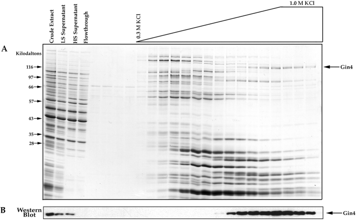
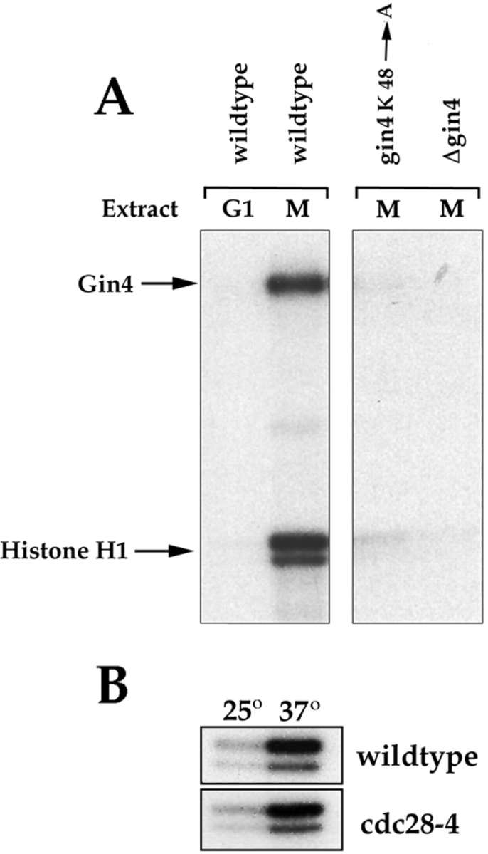
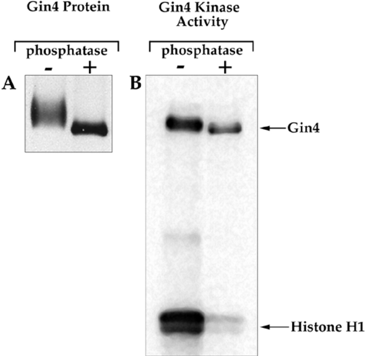
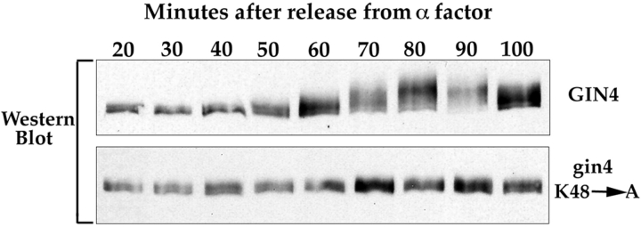
Little is known about the pathways used by cyclins and cyclin-dependent kinases to induce the events of the cell cycle. In budding yeast, a protein called Nap1 binds to the mitotic cyclin Clb2, and Nap1 is required for the ability of Clb2 to induce specific mitotic events, but the role played by Nap1 is unclear. We have used genetic and biochemical approaches to identify additional proteins that function with Nap1 in the control of mitotic events. These approaches have both identified a protein kinase called Gin4 that is required for the ability of Clb2 and Nap1 to promote the switch from polar to isotropic bud growth that normally occurs during mitosis. Gin4 is also required for the ability of Clb2 and Nap1 to promote normal progression through mitosis. The Gin4 protein becomes phosphorylated as cells enter mitosis, resulting in the activation of Gin4 kinase activity, and the phosphorylation of Gin4 is dependent upon Nap1 and Clb2 in vivo. Affinity chromatography experiments demonstrate that Gin4 binds tightly to Nap1, indicating that the functions of these two proteins are closely tied within the cell. These results demonstrate that the activation of Gin4 is under the control of Clb2 and Nap1, and they provide an important step towards elucidating the molecular pathways that link cyclin-dependent kinases to the events they control.


Figures

Figure 3
Figure 3
Deletion of the GIN4 gene causes a prolonged mitotic arrest in cells that are dependent upon Clb2 for survival. Cells were grown overnight at 30°C until they reached an OD of 0.5 (control cells) or 0.8 (Δgin4, Δclb1,3,4, Δbar1 cells), and α-factor was then added to 2 μg/ml at t = 0. At each time point, 1.5 ml of culture was removed and analyzed for Clb2 levels or for the presence of mitotic spindles, as previously described (Kellogg and Murray, 1995; Pringle et al., 1991). The strains used in this experiment carry deletions of the BAR1 gene to prevent them from breaking through the α-factor arrest. (A) A plot of the percentage of cells with a mitotic spindle as a function of time after the addition of α-factor to log phase cultures of a Δgin4, Δclb1,3,4, Δbar1 strain and a Δclb1,3,4, Δbar1 control strain. The percentage of cells with mitotic spindles was determined by counting spindles in random fields of cells. Over 200 cells were counted for each data point. After 3 h, the spindles began to appear unusually thick and bent, and it became difficult to accurately count the number of cells with spindles. (B) A Western blot showing the amount of Clb2 present as a function of time after the addition of α-factor to log phase cultures of the same strains shown in A. (C) Examples of the short spindles observed in the Δgin4, Δclb1,3,4, Δbar1 strain 2 h after addition of α-factor to a log phase culture.
Figure 5
Figure 5
Affinity purification of Nap1-binding proteins. (A) Crude extracts from log phase yeast cells were loaded onto a Nap1 affinity column. After washing with buffer, the column was eluted with a gradient of KCl, and samples from each fraction were precipitated with TCA and loaded onto a 12.5% SDS-polyacrylamide gel. The first few fractions before the start of the salt gradient show the final fractions of the wash. The gel is stained with Coomassie blue. (B) The same fractions shown in A were loaded onto a 10% SDS-polyacrylamide gel and transferred to nitrocellulose, which was then probed with an anti-Gin4 antibody. For the affinity column elution fractions we loaded 1/10 the amount of protein that was loaded onto the gel shown in A.
Figure 7
Figure 7
Gin4 from mitotic cells phosphorylates histone H1 and undergoes autophosphorylation in vitro. (A) Cells from the indicated strains were arrested in G1 with α-factor or in mitosis with benomyl. Extracts were then made from the arrested cells and Gin4 was immunoprecipitated and assayed for kinase activity using histone H1 as a substrate. (B) A control showing that the kinase activity present in Gin4 immunoprecipitates is not due to Cdc28. Wild-type or cdc28-4 cells were grown at 22°C and arrested in mitosis with benomyl. Gin4 was then immunoprecipitated from the arrested cells and kinase activity was measured either at 22°C or 37°C.
Figure 9
Figure 9
The kinase activity of Gin4 is activated by phosphorylation. (A) The Gin4 protein was immunoprecipitated from mitotic cells and then split into two samples. One sample was treated with lambda phosphatase (New England Biolabs Inc.), while the other sample was treated identically, but with no added phosphatase. The precipitated protein was then resolved on a 9% SDS-polyacrylamide gel and detected by Western blotting. (B) The Gin4 protein was immunoprecipitated and treated with lambda phosphatase as in A. The immunoprecipitate was then washed with kinase buffer several times and assayed for kinase activity.
Figure 10
Figure 10
The Gin4 protein appears to undergo autophosphorylation in vivo. gin4K48A, Δclb1,3,4 cells and Δclb1,3,4 control cells were released from an α-factor arrest, and samples were taken at the indicated time points and probed for the Gin4 protein by Western blotting.
Figure 11
Figure 11
Expression of Clb2 in cells arrested in interphase leads to phosphorylation of Gin4. A bar1 strain carrying an integrated copy of Clb2Δ176 under the control of the gal1 promoter was grown in raffinose and arrested in interphase by incubation in the presence of α-factor for 3 h. The culture was then divided in half, and Clb2Δ176 expression was induced in one half by transferring the cells to media containing galactose and α-factor. At the times indicated, samples were taken from each culture and used for an anti-Gin4 Western blot.
Figure 1
Figure 1
An example of the cellular morphology observed for the ecm1 complementation group.
Figure 2
Figure 2
Deletion of the GIN4 gene causes cells to have an elongated bud morphology. Cells were grown to log phase in YPD liquid media at 30°C, and were photographed using a ×100 objective with Nomarski optics. The genotype of each strain is indicated above each picture.
Figure 4
Figure 4
Gin4 is not required for the activation of cyclin-dependent kinase activity by Clb2. (A) A time course showing the appearance of Clb2-associated kinase activity during mitosis. Wild-type and Δgin4 cells were released from an α-factor arrest and then assayed for Clb2-associated kinase activity during the cell cycle as previously described (Kellogg and Murray, 1995). (B) The same samples used for the kinase assay in A were probed for Clb2 levels by Western blotting as previously described (Kellogg and Murray, 1995).
Figure 6
Figure 6
Nap1 coprecipitates with Gin4 in crude cell extracts. Crude cell extracts were made from either wild-type cells or Δgin4 control cells, and the extracts were then used for Gin4 immunoprecipitations. The Gin4 immunoprecipitates were washed either with buffer containing 0.4 M NaCl or 1.0 M NaCl, and were then probed with anti-Nap1 antibody to detect coprecipitated Nap1.
Figure 8
Figure 8
The Gin4 kinase is activated and phosphorylated during mitosis in a Nap1-dependent manner. (A) Δclb1,3,4, Δbar1 cells and Δnap1, Δclb1,3,4, Δbar1 cells were released from an α-factor arrest, and samples were taken at the indicated time points and assayed for Gin4-associated kinase activity using histone H1 as a substrate. (B) The same samples shown in A were assayed for Clb2-associated kinase activity. (C) The same samples shown in A were probed for the Clb2 protein by Western blotting. (D) The same samples shown in A were probed for the Gin4 protein by Western blotting.

References

    1. Amon A, Tyers M, Futcher B, Nasmyth K. Mechanisms that help the yeast cell cycle clock tick: G2 cyclins transcriptionally activate G2 cyclins and repress G1 cyclins. Cell. 1993;74:993–1007. - PubMed
    1. Amon A, Irniger S, Nasmyth K. Closing the cell cycle circle in yeast: G2 cyclin proteolysis initiated at mitosis persists until the activation of G1 cyclins in the next cycle. Cell. 1994;77:1037–1050. - PubMed
    1. Anderson CW, Baum PR, Gesteland RF. Processing of adenovirus 2-induced proteins. J Virol. 1973;12:241–252. - PMC - PubMed
    1. Fitch I, Dahmann C, Surana U, Amon A, Nasmyth K, Goetsch L, Byers B, Futcher B. Characterization of four B-type cyclin genes of the budding yeast Saccharomyces cerevisiae. . Mol Biol Cell. 1992;3:805–818. - PMC - PubMed
    1. Ghiara JB, Richardson HE, Sugimoto K, Henze M, Lew DJ, Witenberg C, Reed SI. A cyclin B homolog in S. cerevisiae: chronic activation of the Cdc28 protein kinase by cyclin prevents exit from mitosis. Cell. 1991;65:163–174. - PubMed

Publication types

MeSH terms

Associated data