. 1997 Jul 21;186(2):239-45.
doi: 10.1084/jem.186.2.239.

Class I-restricted cross-presentation of exogenous self-antigens leads to deletion of autoreactive CD8(+) T cells

Affiliations

Class I-restricted cross-presentation of exogenous self-antigens leads to deletion of autoreactive CD8(+) T cells

C Kurts et al. J Exp Med. .

Abstract

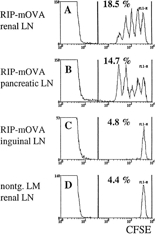
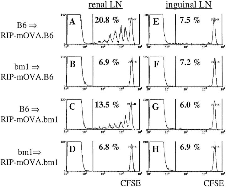
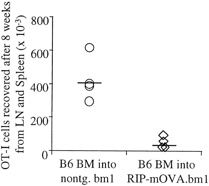
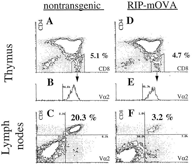
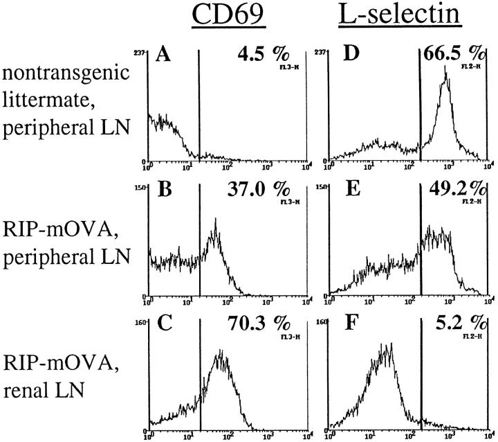
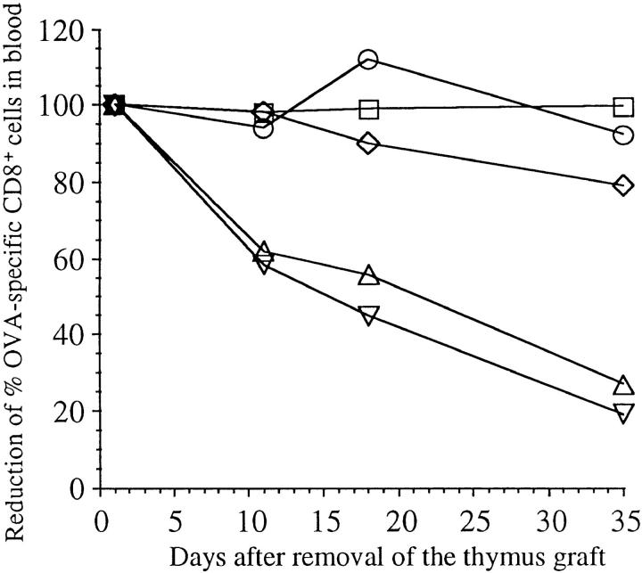
In this report, we show that cross-presentation of self-antigens can lead to the peripheral deletion of autoreactive CD8(+) T cells. We had previously shown that transfer of ovalbumin (OVA)-specific CD8(+) T cells (OT-I cells) into rat insulin promoter-membrane-bound form of OVA transgenic mice, which express the model autoantigen OVA in the proximal tubular cells of the kidneys, the beta cells of the pancreas, the thymus, and the testis of male mice, led to the activation of OT-I cells in the draining lymph nodes. This was due to class I-restricted cross-presentation of exogenous OVA on a bone marrow-derived antigen presenting cell (APC) population. Here, we show that adoptively transferred or thymically derived OT-I cells activated by cross-presentation are deleted from the peripheral pool of recirculating lymphocytes. Such deletion only required antigen recognition on a bone marrow-derived population, suggesting that cells of the professional APC class may be tolerogenic under these circumstances. Our results provide a mechanism by which the immune system can induce CD8(+) T cell tolerance to autoantigens that are expressed outside the recirculation pathway of naive T cells.


Figures

Figure 1
Figure 1
Proliferation of OT-I cells in lymph nodes draining OVA-expressing tissues of RIP–mOVA mice. 5 × 10⁶ CFSE-labeled OT-I cells were injected intravenously into RIP–mOVA mice and nontransgenic littermates. 43 h later, lymphocytes prepared from the renal (A), pancreatic (B), and inguinal (C) LN of RIP–mOVA mice, and from the renal LN of nontransgenic recipients were analyzed by flow cytometry. Profiles were gated on CD8+ cells.
Figure 2
Figure 2
Bone marrow–derived APC activate OT-I cells in the draining LN of RIP–mOVA mice. 5 × 10⁶ CFSE labeled OT-I cells were transferred into RIP–mOVA mice backcrossed to B6 (A, B, E, F) or bm1 (C, D, G, H) and grafted with B6 (A, C, E, G) or bm1 (B, D, F, H) bone marrow. After 3 d, renal (A–D) and inguinal (E–H) lymph node cells were examined by flow cytometry. Profiles were gated for CD8+ cells.
Figure 3
Figure 3
OT-I cells are deleted in response to recognition of antigen on cross-presenting APC. Bone marrow from B6 mice was grafted into RIP–mOVA.bm1 mice and nontransgenic littermates. 14 wk later, 6 × 10⁶ OT-I cells were adoptively transferred, and after 8 wk the number of Vα2+Vβ5+CD8+ cells in the LN and spleen of the recipients was determined by flow cytometry. The proportion of Vα2+Vβ5+ cells in the CD8+ population was 7.5–10% in nontransgenic, and 1.4–3.5% in transgenic recipients. An average of 1.4% of CD8+ cells were Vα2+Vβ5+ in uninjected mice. To derive the total number of OT-I cells, this 1.4% was subtracted from the proportion of Vα2+Vβ5+ cells in the CD8+ cells in experimental mice and the difference was multiplied with the proportion of CD8+ T cells in the live cells and with the number of live cells. These results are representative for two such experiments consisting of four mice per group.
Figure 4
Figure 4
Peripheral deletion of OT-I cells that mature in the thymus of TG-RIP mice. Thymus grafts from TG-littermate mice (A and B) and TG-RIP mice (D and E) were analyzed for CD4+, CD8+, and Vα2+ cells by flow cytometry 16 wk after bone marrow reconstitution. Expression of Vα2 by CD4CD8+ cells is shown in the histograms B and E. LN cells of the same transgenic (C) and nontransgenic (F) mice were stained for CD8+ and Vα2+ expression. The same staining conditions for Vα2 were used for thymus and LN cells. The data shown here is representative for eight pairs of manipulated mice investigated.
Figure 5
Figure 5
Analysis of the activation status of OT-I cells in TG-RIP mice. Expression of CD69 (A–C) and L-selectin (D–F) by Vα2+ lymphocytes from the peripheral LN of TG-nontransgenic mice (A and D) and the peripheral (B and E) and renal (C and F) LN of TG-RIP mice.
Figure 6
Figure 6
Disappearance of OT-I cells in TG-RIP mice after removal of their thymus graft. Thymus graft was removed from TG-RIP mice and TG-nontransgenic controls 3 mo after implantation. The proportion of CD8+ peripheral blood cells that were Vα2+Vβ5+ was then determined by flow cytometry on days 2, 11, 18, and 35 after thymectomy. The proportion found at day 2 after removal of the thymus grafts was considered 100% in this particular mouse, and the following values were given as a percent of this starting value. TG-RIP, not operated (○); TG-RIP, thymus graft removed (▿); TG-RIP, thymus graft and left kidney removed (▵); TG-nontransgenic littermate, not operated (□); TG-nontransgenic littermate, thymus graft removed (⋄).

References

    1. Wooley PH, Luthra HS, Stuart JM, David CS. Type II collagen-induced arthritis in mice. I. Major histocompatibility complex (I region) linkage and antibody correlates. J Exp Med. 1981;154:688–700. - PMC - PubMed
    1. Zamvil SS, Steinman L. The T lymphocyte in experimental allergic encephalomyelitis. Annu Rev Immunol. 1990;8:579–621. - PubMed
    1. Ohashi PS, Oehen S, Buerki K, Pircher H, Ohashi CT, Odermatt B, Malissen B, Zinkernagel RM, Hengartner H. Ablation of “tolerance” and induction of diabetes by virus infection in viral antigen transgenic mice. Cell. 1991;65:305–317. - PubMed
    1. Oldstone MB, Nerenberg M, Southern P, Price J, Lewicki H. Virus infection triggers insulin-dependent diabetes mellitus in a transgenic model: role of anti-self (virus) immune response. Cell. 1991;65:319–331. - PubMed
    1. Schonrich G, Kalinke U, Momburg F, Malissen M, Schmitt-Verhulst AM, Malissen B, Hammerling GJ, Arnold B. Down-regulation of T cell receptors on self-reactive T cells as a novel mechanism for extrathymic tolerance induction. Cell. 1991;65:293–304. - PubMed

Publication types

Substances