. 1997 Jul 21;186(2):307-12.
doi: 10.1084/jem.186.2.307.

Myelin basic protein-specific T helper 2 (Th2) cells cause experimental autoimmune encephalomyelitis in immunodeficient hosts rather than protect them from the disease

Affiliations

Myelin basic protein-specific T helper 2 (Th2) cells cause experimental autoimmune encephalomyelitis in immunodeficient hosts rather than protect them from the disease

J J Lafaille et al. J Exp Med. .

Abstract

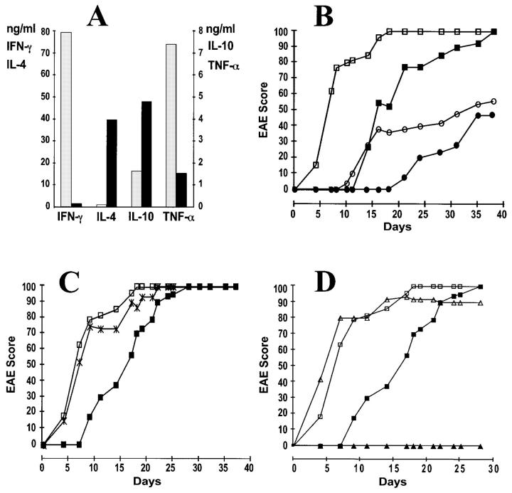
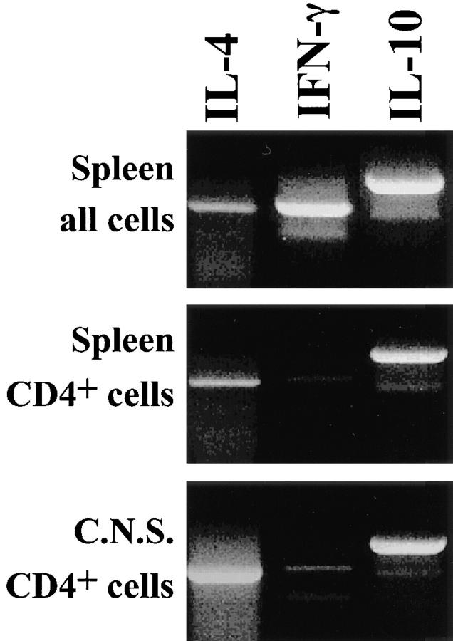
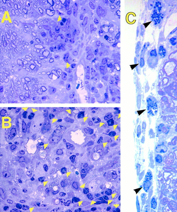
Chronic inflammatory autoimmune diseases such as multiple sclerosis, diabetes, and rheumatoid arthritis are caused by CD4(+) Th1 cells. Because Th2 cells antagonize Th1 cell functions in several ways, it is believed that immune deviation towards Th2 can prevent or cure autoimmune diseases. Experimental autoimmune encephalomyelitis (EAE) is a demyelinating disease used as a model for multiple sclerosis. Using an adoptive transfer system we assessed the role of Th1 and Th2 cells in EAE. In vitro generated Th1 and Th2 cells from myelin basic protein (MBP)-specific TCR transgenic mice were transferred into normal and immunodeficient mice. Th1 cells caused EAE in all recipients after a brief preclinical phase. Surprisingly, Th2 cells also caused EAE in RAG-1 KO mice and in alphabeta T cell-deficient mice, albeit after a longer preclinical phase. Normal or gammadelta T cell-deficient mice were resistant to EAE induced by Th2 cells. The histopathological features of this disease resembled those of an allergic process. In addition, disease induction by Th1 cells was not altered by coadmininstration of Th2 cells in any of the recipients. These findings indicate that MBP-specific Th2 cells have the potential to induce EAE and that the disease induced by previously activated Th1 cells cannot be prevented by normal lymphocytes nor by previously activated Th2 cells.


Figures

Figure 1
Figure 1
(A) Interleukins in the supernatants of MBP-specific Th1 and Th2 cultures at the day of injection. (Gray bars) Th1 cultures; (black bars) Th2 cultures. (B) Both Th1 and Th2 anti-MBP T cells induce EAE in RAG-1 KO recipients. Mice were injected intravenously with 5 × 10⁶ Th1 cells (open squares, n = 9), 5 × 10⁶ Th2 cells (filled squares, n = 6), 0.2 × 10⁶ Th1 cells (open circles, n = 5), or 0.2 × 10⁶ Th2 cells (filled circles, n = 3). Data is presented as a percentage of maximum possible EAE that would be reached if all the mice were at level 5. Animals reaching level 5 were sacrificed and their score was kept at level 5 for the remaining of the experiment. Data from one representative experiment (out of five) is shown. (C) Coinjection of Th2 does not alter the kinetics of EAE caused by Th1 cells. RAG-1 KO recipient mice were injected intravenously with 10 × 106 Th1 cells (open squares, n = 12), 10 × 106 Th2 cells (filled squares, n = 12), or a mixture of 5 × 10⁶ Th1 cells and 5 × 10⁶ Th2 cells (cross, n = 4). (D) MBP-specific Th2 cells do not induce EAE in syngeneic immunocompetent recipients. Ten million Th1 cells (open symbols) or Th2 cells (filled symbols) were injected intravenously into RAG-1 recipients (squares, n = 12 for both Th1 or Th2 recipients) or B10.PL recipients (triangles, n = 6 for both Th1 and Th2 recipients).
Figure 2
Figure 2
Th2 cells but not Th1 cells are recovered from animals to which cells from Th2 cell cultures had been injected. Spleen (top two panels) and CNS cells from mice injected with Th2 cells were prepared 40 d after injection, when mice were at level 2 and 3 of EAE. The lower two panels show purified CD4-positive cells. RT-PCR was performed to determine mRNA levels of IL-4, IFN-γ, and IL-10. The IFN-γ mRNA present in total splenocytes and absent in CD4 purified splenocytes and lymphocytes from CNS is likely coming from NK cells.
Figure 3
Figure 3
Unusually high numbers of polymorphonuclear cells and mast cells are found in spinal cord lesions of Th2 cell– but not in Th1 cell–induced EAE. 1-μm thin Epoxy sections from mice with EAE (clinical grade 2) injected with Th1 (A) or Th2 (B and C) MBP-specific T cells were stained with toluidine blue. (Yellow arrows) Polymorphonuclear cells in the white matter infiltrates. (Black arrows) Mast cells in the subarachnoid space of the meninges. Original magnification: (A and B) ×1,500; (C) ×3,300.
Figure 4
Figure 4
Th1 cells do not migrate faster into the CNS parenchyma than Th2 cells. Th1 (gray bars) and Th2 cells (black bars) were labeled with 51Cr and injected intravenously. After 45 and 90 h the mice were perfused and the radioactivity of several organs was determined in a gamma counter. At all time points most of the counts were found in the liver. Data are presented as a ratio between counts in the CNS and counts in the liver. One representative experiment (of three) is shown.

References

    1. Mosmann TR, Coffman RL. Th1 and Th2 cells: different patterns of lymphokine secretion lead to different functional properties. Ann Rev Immunol. 1989;7:145–173. - PubMed
    1. Abbas AK, Murphy KM, Sher A. Functional diversity of helper lymphocytes. Nature (Lond) 1996;383:787–793. - PubMed
    1. Seder RA, Paul WE, Davis MM, Fazekas de St B, Groth The presence of interleukin-4 during in vitro priming determines the lymphokine-producing potential of CD4+T cells from T cell receptor transgenic mice. J Exp Med. 1992;176:1091–1098. - PMC - PubMed
    1. Hsieh CS, Heimberger AB, Gold JS, O'Garra A, Murphy KM. Differential regulation of T helper phenotype development by interleukins 4 and 10 in an αβ T-cell receptor transgenic system. Proc Natl Acad Sci USA. 1992;89:6065–6069. - PMC - PubMed
    1. Hsieh CS, Macatonia SE, Tripp CS, Wolf SF, O'Garra A, Murphy KM. Development of Th1 CD4+T cells through IL-12 produced by Listeria-induced macrophages. Science (Wash DC) 1993;260:547–549. - PubMed

Publication types

Substances