. 1997 Jul 21;186(2):331-6.
doi: 10.1084/jem.186.2.331.

Distinct roles for signals relayed through the common cytokine receptor gamma chain and interleukin 7 receptor alpha chain in natural T cell development

Affiliations

Distinct roles for signals relayed through the common cytokine receptor gamma chain and interleukin 7 receptor alpha chain in natural T cell development

A Boesteanu et al. J Exp Med. .

Abstract

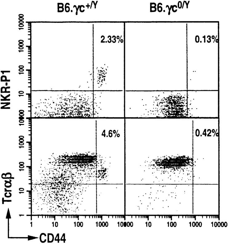
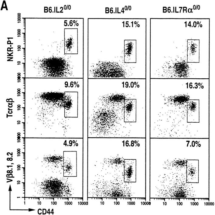
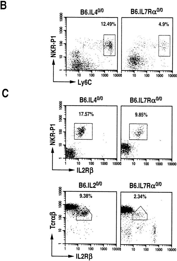
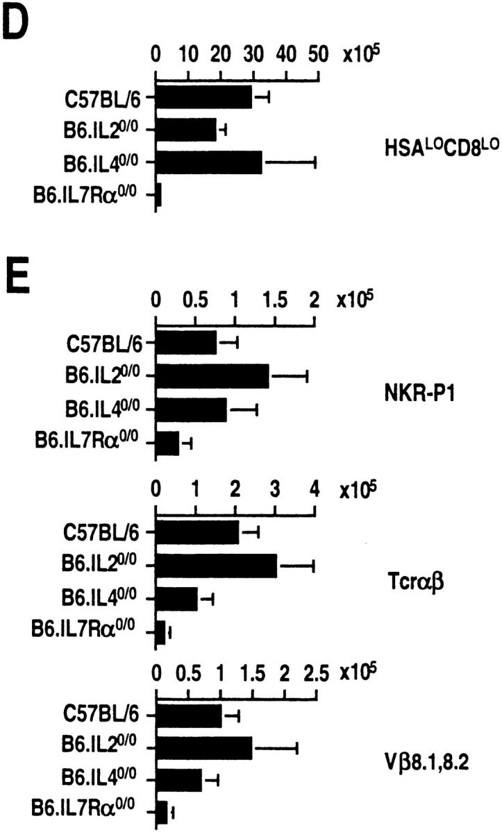
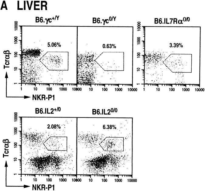
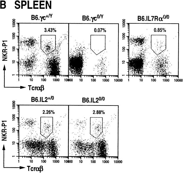
The commitment, differentiation, and expansion of mainstream alpha/beta T cells during ontogeny depend on the highly controlled interplay of signals relayed by cytokines through their receptors on progenitor cells. The role of cytokines in the development of natural killer (NK)1(+) natural T cells is less clearly understood. In an approach to define the role of cytokines in the commitment, differentiation, and expansion of NK1(+) T cells, their development was studied in common cytokine receptor gamma chain (gammac) and interleukin (IL)-7 receptor alpha (IL-7Ralpha)-deficient mice. These mutations block mainstream alpha/beta T cell ontogeny at an early prethymocyte stage. Natural T cells do not develop in gammac-deficient mice; they are absent in the thymus and peripheral lymphoid organs such as the liver and the spleen. In contrast, NK1(+) T cells develop in IL-7Ralpha-deficient mice in the thymus, and they are present in the liver and in the spleen. However, the absolute number of NK1(+) T cells in the thymus of IL-7Ralpha-deficient mice is reduced to approximately 10%, compared to natural T cell number in the wild-type thymus. Additional data revealed that NK1(+) T cell ontogeny is not impaired in IL-2- or IL-4-deficient mice, suggesting that neither IL-2, IL-4, nor IL-7 are required for their development. From these data, we conclude that commitment and/or differentiation to the NK1(+) natural T cell lineage requires signal transduction through the gammac, and once committed, their expansion requires signals relayed through the IL-7Ralpha.


Figures

Figure 1
Figure 1
Common cytokine receptor γc-deficient mice do not develop thymic NTα/β cells. Dot plots displaying CD44+ NKR-P1+ and CD44+TCR-α/β+ T cells among HSAlow CD8low thymocytes of B6.γc0/Y (seventh generation backcross to C57BL/6) and B6.IL-2Rγc+/Y littermates. HSAlowCD8low thymocyte population was electronically gated and CD44highNKR-P1+ T cells were analyzed with a FACScan® flow cytometer.
Figure 2
Figure 2
IL-2–, IL-4–, and IL-7Rα–deficient mice develop thymic NTα/β T cells. (A) Dot plots of CD44highNKR-P1+, CD44highTCR-α/βmed, and CD44highTCR-β8.1,8.2med T cells among HSAlowCD8low thymocytes of B6.IL-20/0 (n = 5), B6.IL-40/0 (n = 6) and B6.IL-7Rα0/0 mice (n = 6). (B) Dot plots of Ly6ChighNKR-P1+ T cells among HSAlowCD8low thymocytes. (C) Dot plots of IL-2 Rβ+NKR-P1+ (B6.IL-40/0 and B6.IL-7Rα0/0) and of IL-2Rβ+TCR-α/β+ (B6.IL-20/0 and B6.IL-7Rα0/0) T cells among HSAlowCD8low thymocytes. In B and C, the percentage of NT cells was almost equal in B6.IL-40/0, about half in B6.IL-7Rα0/0, or up to twofold greater in B6.IL-20/0 compared with those in the wild type. (D) Absolute numbers of HSAlowCD8low thymocytes calculated as the thymocyte number times the fraction of this subset. (E) Thymic NT cells in wild-type, IL-20/0, IL-40/0 and IL-7Rα0/0 mice. NT cell number was calculated from the percentages of the double-positive CD44+ and NKR-P1+, TCR-α/β+ or Vβ8.1,8.2+ thymocytes within the electronically gated HSAlowCD8low population in D as described previously (5).
Figure 2
Figure 2
IL-2–, IL-4–, and IL-7Rα–deficient mice develop thymic NTα/β T cells. (A) Dot plots of CD44highNKR-P1+, CD44highTCR-α/βmed, and CD44highTCR-β8.1,8.2med T cells among HSAlowCD8low thymocytes of B6.IL-20/0 (n = 5), B6.IL-40/0 (n = 6) and B6.IL-7Rα0/0 mice (n = 6). (B) Dot plots of Ly6ChighNKR-P1+ T cells among HSAlowCD8low thymocytes. (C) Dot plots of IL-2 Rβ+NKR-P1+ (B6.IL-40/0 and B6.IL-7Rα0/0) and of IL-2Rβ+TCR-α/β+ (B6.IL-20/0 and B6.IL-7Rα0/0) T cells among HSAlowCD8low thymocytes. In B and C, the percentage of NT cells was almost equal in B6.IL-40/0, about half in B6.IL-7Rα0/0, or up to twofold greater in B6.IL-20/0 compared with those in the wild type. (D) Absolute numbers of HSAlowCD8low thymocytes calculated as the thymocyte number times the fraction of this subset. (E) Thymic NT cells in wild-type, IL-20/0, IL-40/0 and IL-7Rα0/0 mice. NT cell number was calculated from the percentages of the double-positive CD44+ and NKR-P1+, TCR-α/β+ or Vβ8.1,8.2+ thymocytes within the electronically gated HSAlowCD8low population in D as described previously (5).
Figure 3
Figure 3
NTα/β cells do not develop in peripheral lymphoid organs of γc-deficient mice, but develop in IL-7Rα0/0 and IL-20/0 animals. Dot plots displaying NKR-P1+TCR-α/β+ cells among mononuclear cells isolated from the liver (A) and spleen (B) of wild-type and mutant mice. Liver NTα/β cells were stained with anti–NKR-P1-PE and anti–TCR-β-biotin (B6.γc0/Y and B6.IL-7Rα0/0) or anti–TCR-β-PE and anti–NKR-P1-biotin (B6.IL-2+/0 and B6.IL-20/0). Splenic NTα/β cells were stained with anti–B220-FITC, anti-TCR-β-PE and anti-NKR-P1-biotin, and identified among B220null splenocytes. In all cases, the biotinylated antibodies were detected by staining with streptavidin-RED670. The staining pattern of intrahepatic NT cells was observed in over 20 different preparations (see also reference 4).
Figure 3
Figure 3
NTα/β cells do not develop in peripheral lymphoid organs of γc-deficient mice, but develop in IL-7Rα0/0 and IL-20/0 animals. Dot plots displaying NKR-P1+TCR-α/β+ cells among mononuclear cells isolated from the liver (A) and spleen (B) of wild-type and mutant mice. Liver NTα/β cells were stained with anti–NKR-P1-PE and anti–TCR-β-biotin (B6.γc0/Y and B6.IL-7Rα0/0) or anti–TCR-β-PE and anti–NKR-P1-biotin (B6.IL-2+/0 and B6.IL-20/0). Splenic NTα/β cells were stained with anti–B220-FITC, anti-TCR-β-PE and anti-NKR-P1-biotin, and identified among B220null splenocytes. In all cases, the biotinylated antibodies were detected by staining with streptavidin-RED670. The staining pattern of intrahepatic NT cells was observed in over 20 different preparations (see also reference 4).

References

    1. Vicari AP, Zlotnik A. Mouse NK1.1+T cells: a new family of T cells. Immunol Today. 1996;17:71–76. - PubMed
    1. Smiley ST, Kaplan MH, Grusby MJ. Immunoglobulin E production in the absence of interleukin-4 secreting CD1 dependent cells. Science (Wash DC) 1997;275:977–979. - PubMed
    1. Chen Y-H, Chiu NM, Mandal M, Wang N, Wang C-R. Impaired NK1+T cell development and early IL-4 production in CD1 deficient mice. Immunity. 1997;6:459–467. - PubMed
    1. Mendiratta SK, Martin WD, Hong S, Boesteanu A, Joyce S, Van Kaer L. CD1d1mutant mice are deficient in natural T cells that promptly produce IL-4. Immunity. 1997;6:469–477. - PubMed
    1. Joyce S, Negishi I, Boesteanu A, DeSilva AD, Sharma P, Chorney MJ, Loh DY, Van Kaer L. Expansion of natural (NK1+) T cells that express αβ T cell receptors in transporters associated with antigen presentation-1 null and thymus leukemia antigen positive mice. J Exp Med. 1996;184:1579–1584. - PMC - PubMed

Publication types