. 1997 Jul 21;186(2):337-42.
doi: 10.1084/jem.186.2.337.

Assembly and regulation of the CD40 receptor complex in human B cells

Affiliations

Assembly and regulation of the CD40 receptor complex in human B cells

M R Kuhné et al. J Exp Med. .

Abstract

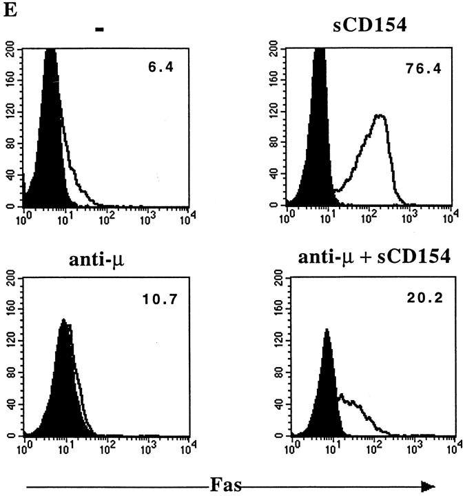
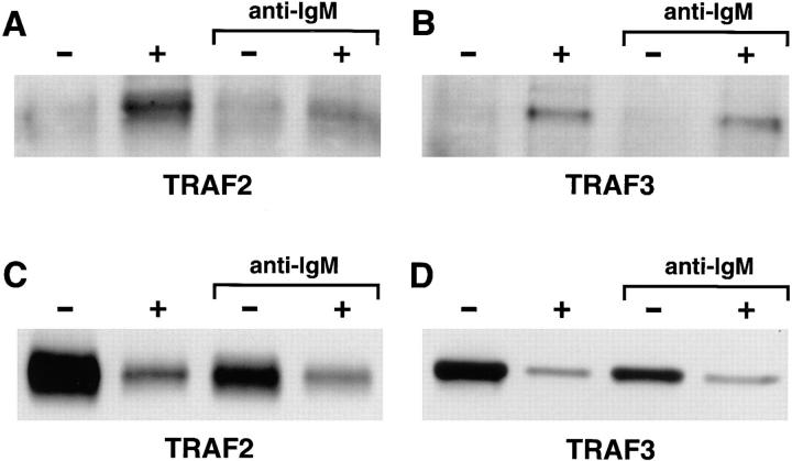
CD40 is a member of the tumor necrosis factor (TNF) receptor superfamily. Studies with human B cells show that the binding of CD154 (gp39, CD40L) to CD40 recruits TNF receptor- associated factor 2 (TRAF2) and TRAF3 to the receptor complex, induces the downregulation of the nonreceptor-associated TRAFs in the cell and induces an increased expression of Fas on the cell surface. Combined signaling through the interluekin 4 receptor and CD40 induces an increased expression of Fas with a commensurate increase in the level of TRAF2, but not TRAF3, that is recruited to the receptor complex. In contrast, engagement of the membrane immunoglobulin and CD40 limits Fas upregulation and reduces the recruitment of TRAF2, relative to TRAF3, to the CD40 receptor complex. These studies show that the TRAF composition of the CD40 receptor complex can be altered by signals that influence B cell differentiation.


Figures

Figure 1
Figure 1
Stimulation with sCD154 induces recruitment of TRAF2 and TRAF3 to CD40. (A) DND39 cells (2 × 10⁷ cells/lane) were either left unstimulated (lanes 1, 2, 5, and 7), or stimulated with sCD154 (4 nM) (lanes 3, 4, 6, and 8) for 15 min before lysis and immunoprecipitation with either irrelevant mouse IgG1 (lanes 1, 3) and or with anti-human CD40 mouse IgG1 monoclonal BE-1 (2, 4) or S2C6 (lanes 5, 6, and 7, 8), respectively. The immunoprecipitated samples were immunoblotted for TRAF2 (arrowhead) (lanes 1–6) or for TRAF3 (indicated by asterisk) (lanes 7, 8). This experiment is representative of 12 experiments.
Figure 3
Figure 3
Anti-μ cross-linking reduces TRAF2 association and Fas upregulation induced by CD40 signaling. DND39 cells (2 × 10⁷ cells/lane) were incubated in media or with 10 μg/ml goat anti–human IgM for 24 h and both groups were either left unstimulated (minus) or stimulated with sCD154 (4 nM) (plus) for 15 min. Lysates were immunoprecipitated for CD40 and immunoblotted for TRAF2 (A) or TRAF3 (B). (C and D) Immunoblot analysis of cytosolic TRAF content after GST–CD40cyt immunoprecipitations. The lysates used for the anti-CD40 precipitations described in A and B were further precipitated with GST–CD40cyt. 5 × 10⁶ cells/lane were loaded for the TRAF2 blot, while the TRAF3 blot received 2 × 10⁷ cell equivalents per lane. (E) Fluorescence-activated cell sorting analysis of DND39 cells after treatment with anti-IgM and sCD154. DND39 cells were incubated for 24 h with media (minus), 10 μg/ml anti-IgM, sCD154, or both (as indicated). Fas expression was detected with anti-CD95 monoclonal antibody (open profile) or a control mouse IgG1 monoclonal (closed profile). The mean fluorescent intensity of the cells is indicated in the upper righthand corner. This experiment is representative of four experiments.
Figure 3
Figure 3
Anti-μ cross-linking reduces TRAF2 association and Fas upregulation induced by CD40 signaling. DND39 cells (2 × 10⁷ cells/lane) were incubated in media or with 10 μg/ml goat anti–human IgM for 24 h and both groups were either left unstimulated (minus) or stimulated with sCD154 (4 nM) (plus) for 15 min. Lysates were immunoprecipitated for CD40 and immunoblotted for TRAF2 (A) or TRAF3 (B). (C and D) Immunoblot analysis of cytosolic TRAF content after GST–CD40cyt immunoprecipitations. The lysates used for the anti-CD40 precipitations described in A and B were further precipitated with GST–CD40cyt. 5 × 10⁶ cells/lane were loaded for the TRAF2 blot, while the TRAF3 blot received 2 × 10⁷ cell equivalents per lane. (E) Fluorescence-activated cell sorting analysis of DND39 cells after treatment with anti-IgM and sCD154. DND39 cells were incubated for 24 h with media (minus), 10 μg/ml anti-IgM, sCD154, or both (as indicated). Fas expression was detected with anti-CD95 monoclonal antibody (open profile) or a control mouse IgG1 monoclonal (closed profile). The mean fluorescent intensity of the cells is indicated in the upper righthand corner. This experiment is representative of four experiments.
Figure 4
Figure 4
Engagement of CD40 results in the loss of immunoprecipitable TRAF2 and TRAF3 from DND39 cells. DND39 cells (2 × 10⁶ cells/ml) were left untreated (minus) or treated with 4 nM sCD154 (plus) for 15 min. After treatment, lysates were immunoprecipitated with either anti-TRAF2 or anti-TRAF3 antibodies. 5 × 10⁶ cell equivalents were loaded per lane.
Figure 2
Figure 2
Pretreatment with IL-4 increases TRAF2, but not TRAF3, recruited to CD40 and increases cell surface Fas expression. DND39 cells (2 × 10⁷ cells/lane) were either left untreated or pretreated for 10 min with human (h)IL-4 (2 ng/ml) (Genzyme, Cambridge, MA); and both groups were either left unstimulated (minus) or stimulated with sCD154 (4 nM) (plus) for 15 min. Lysates were immunoprecipitated for CD40 and immunoblotted for TRAF2 (A) or TRAF3 (B). (C) Fluorescence-activated cell sorting analysis of DND39 cells after treatment with hIL-4 and sCD154. DND39 cells were incubated for 24 h with media (minus), 2 ng/ml hIL-4, sCD154, or both (as indicated). Fas expression was detected with anti-CD95 monoclonal antibody (open profile) or a control mouse IgG1 monoclonal (closed profile). The mean fluorescent intensity of the cells is indicated in the upper righthand corner. This experiment is representative of four experiments.
Figure 2
Figure 2
Pretreatment with IL-4 increases TRAF2, but not TRAF3, recruited to CD40 and increases cell surface Fas expression. DND39 cells (2 × 10⁷ cells/lane) were either left untreated or pretreated for 10 min with human (h)IL-4 (2 ng/ml) (Genzyme, Cambridge, MA); and both groups were either left unstimulated (minus) or stimulated with sCD154 (4 nM) (plus) for 15 min. Lysates were immunoprecipitated for CD40 and immunoblotted for TRAF2 (A) or TRAF3 (B). (C) Fluorescence-activated cell sorting analysis of DND39 cells after treatment with hIL-4 and sCD154. DND39 cells were incubated for 24 h with media (minus), 2 ng/ml hIL-4, sCD154, or both (as indicated). Fas expression was detected with anti-CD95 monoclonal antibody (open profile) or a control mouse IgG1 monoclonal (closed profile). The mean fluorescent intensity of the cells is indicated in the upper righthand corner. This experiment is representative of four experiments.

References

    1. Foy T, Aruffo A, Bajorath J, Buhlmann JE, Noelle RJ. Immune regulation by CD40 and its ligand gp39. Annu Rev Immunol. 1996;14:591–617. - PubMed
    1. Banchereau J, Bazan F, Blanchard D, Briere F, Galizzi JP, van Kooten C, Liu YJ, Rousset F, Saeland S. The CD40 antigen and its ligand. Annu Rev Immunol. 1994;12:881–922. - PubMed
    1. Grewal IS, Xu J, Flavell RA. Impairment of antigen-specific T-cell priming in mice lacking CD40 ligand. Nature (Lond) 1995;378:617–620. - PubMed
    1. Campbell KA, Ovendale PJ, Kennedy MK, Fanslow WC, Reed SG, Maliszewski CR. CD40 ligand is required for protective cell-mediated immunity to Leishmania major. Immunity. 1996;4:283–289. - PubMed
    1. Kawabe T, Naka T, Yoshida K, Tanaka T, Fujiwara H, Suematsu S, Yoshida N, Kishimoto T, Kikutani H. The immune responses in CD40-deficient mice: impaired immunoglobulin class switching and germinal center formation. Immunity. 1994;1:167–178. - PubMed

Publication types