. 1997 Aug 1;17(15):5843-57.
doi: 10.1523/JNEUROSCI.17-15-05843.1997.

Caenorhabditis elegans levamisole resistance genes lev-1, unc-29, and unc-38 encode functional nicotinic acetylcholine receptor subunits

Affiliations

Caenorhabditis elegans levamisole resistance genes lev-1, unc-29, and unc-38 encode functional nicotinic acetylcholine receptor subunits

J T Fleming et al. J Neurosci. .

Abstract

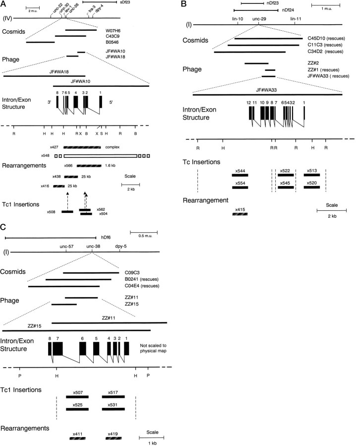
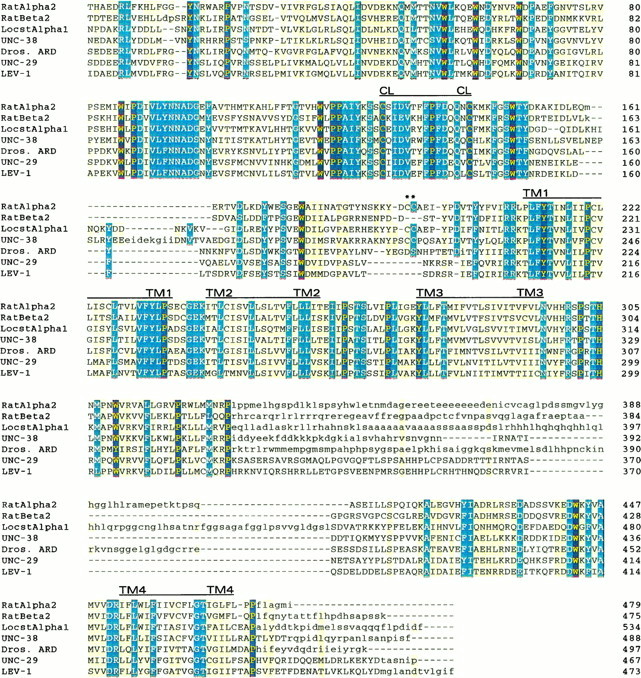
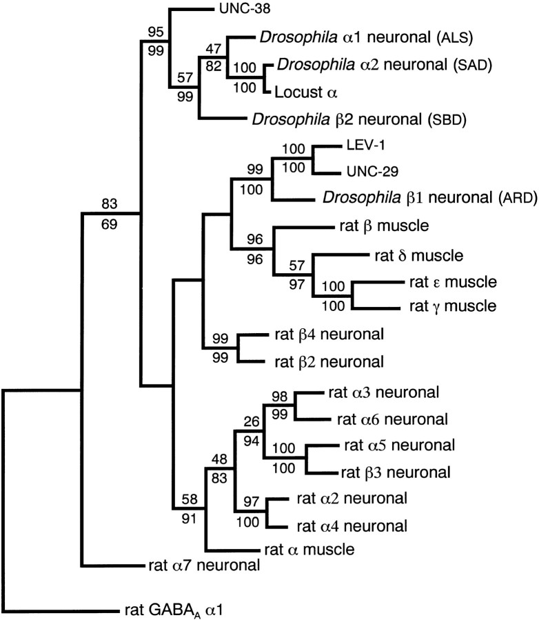
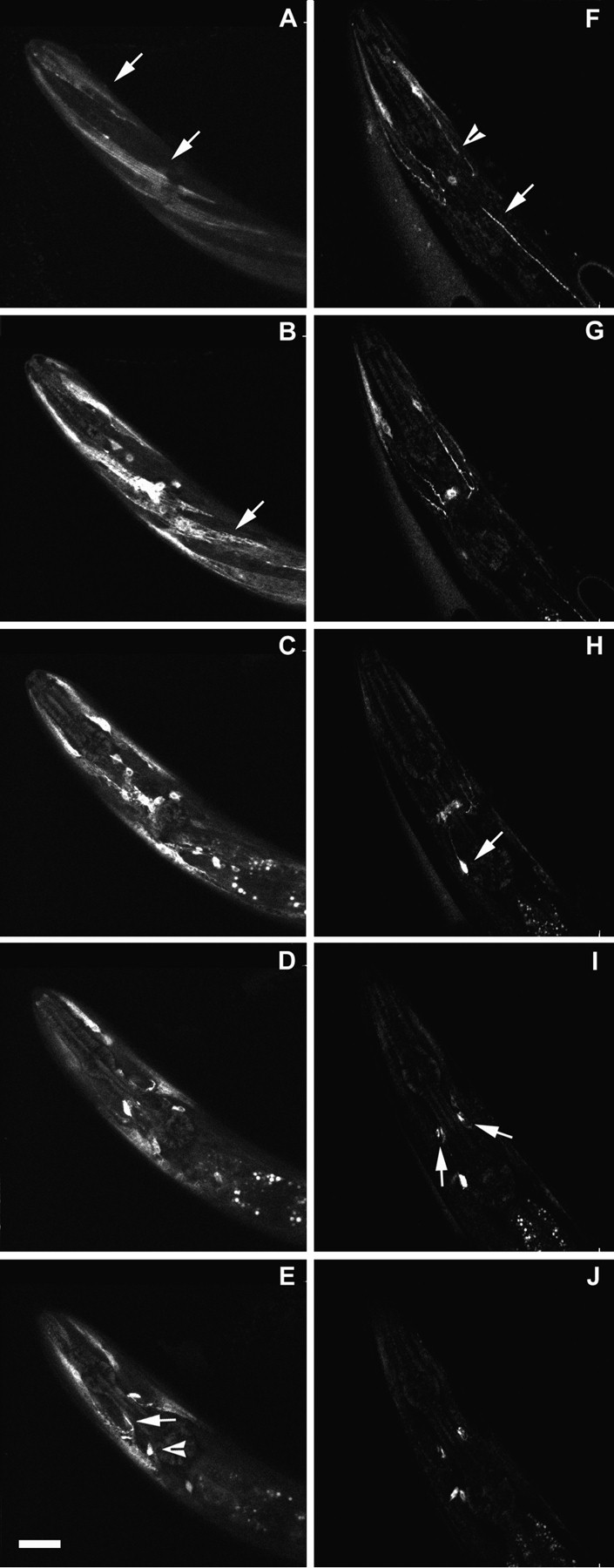
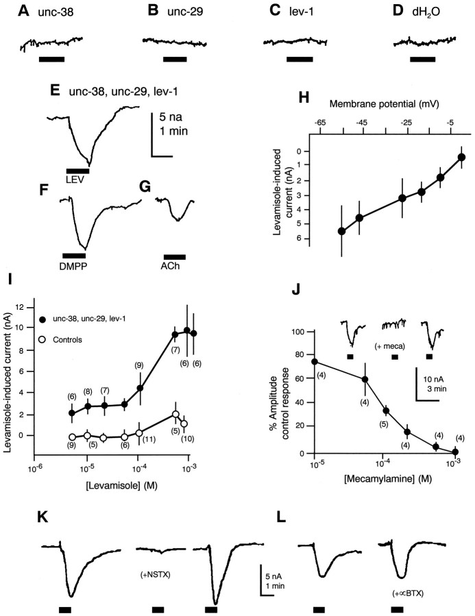
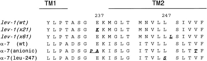
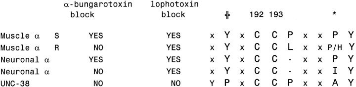
We show that three of the eleven genes of the nematode Caenorhabditis elegans that mediate resistance to the nematocide levamisole and to other cholinergic agonists encode nicotinic acetylcholine receptor (nAChR) subunits. unc-38 encodes an alpha subunit while lev-1 and unc-29 encode non-alpha subunits. The nematode nAChR subunits show conservation of many mammalian nAChR sequence features, implying an ancient evolutionary origin of nAChR proteins. Expression in Xenopus oocytes of combinations of these subunits that include the unc-38 alpha subunit results in levamisole-induced currents that are suppressed by the nAChR antagonists mecamylamine, neosurugatoxin, and d-tubocurarine but not alpha-bungarotoxin. The mutant phenotypes reveal that unc-38 and unc-29 subunits are necessary for nAChR function, whereas the lev-1 subunit is not. An UNC-29-GFP fusion shows that UNC-29 is expressed in body and head muscles. Two dominant mutations of lev-1 result in a single amino acid substitution or addition in or near transmembrane domain 2, a region important to ion channel conductance and desensitization. The identification of viable nAChR mutants in C. elegans provides an advantageous system in which receptor expression and synaptic targeting can be manipulated and studied in vivo.


Figures

Fig. 1.
Fig. 1.
Structures of thelev-1, unc-29, and unc-38nAChR subunit genes. A, lev-1 structure. The position of lev-1 on the genetic map of chromosome IV is shown in relation to nearby genetic markers. The relative contig positions on the physical map of cosmids W07H6, C43C9, and B0564 and λ phage clones JF#WA10 and JF#WA18 derived from thelev-1 region are indicated. The genomic organization of the lev-1 gene is shown. Restriction enzyme sites are indicated to define possible limits of mutations: R,EcoRI; S, SalI;H, HindIII; X,XhoI; and B, BamHI. Mutant alleles found to have substantial DNA sequence rearrangements or Tc1 insertions are diagrammed. The open bar forx548 represents a deletion with a range of end points indicated. x416, x427, x438, andx566 represent complex rearrangements, mostly insertions of the sizes indicated, affecting the restriction fragments spanned by the bar shown for each mutation. Forx504, x508, and x562, the positions of 1.6 kb Tc1 insertions are indicated byarrows. For e211, e289,x21, x38, x61, x400, andx505, no DNA differences were detected. GenBank entryX98601 gives the DNA sequence of lev-1. B,unc-29 structure. The position of unc-29on the genetic map of chromosome I is shown in relation to nearby genetic markers. The relative contig positions on the physical map of cosmids C45D10, C11C3, and C34D2 and λ phage clones ZZ#1, ZZ#2, and JF#WA33 derived from the unc-29 region are indicated. The genomic organization of the unc-29 gene is shown. Restriction enzyme sites: R, EcoRI;S, SalI; and H,HindIII. The positions of a mutation caused by DNA rearrangement and of six mutations caused by apparent transposon insertion are shown relative to the EcoRI fragments that span the unc-29 gene. The sizes of the apparent inserts found were 1.6 kb each for x513, x522,x544, x545, and x554 and 2.5 kb for x520. The inserts of x520,x544, and x554 were lost in revertants, and the insert of x513 hybridized to Tc1. The exact nature and extent of the x415 mutation are unknown, but at least several hundred bases near the 3′ end of the coding seem to be involved (see Materials and Methods). For x401,x417, x429, and x433, noDNA differences were detected. The DNA sequence ofunc-29 is given by GenBank entry U81144.C, Structure of the unc-38 nAChR α subunit gene. The position of unc-38 on the genetic map of chromosome I is shown in relation to nearby genetic markers. The relative contig positions on the physical map of cosmids C09C3, B0241, and C04E4 and λ phage clones ZZ#11 and ZZ#15 are indicated. The genomic organization of the unc-38 gene is shown. Restriction enzyme sites: P, PstI; andH, HindIII. Mutations associated with Tc1 insertion or DNA rearrangements within the HindIII fragment spanning the unc-38 gene are indicated. Forx402, x404, x414, andx511, no DNA differences were found in this fragment. Other than causing a size alteration of the 3.2 kbHindIII fragment, the exact nature and extent of thex411 and x419 mutations are unknown. The DNA sequence of unc-38 is given by GenBank entryX98599.
Fig. 2.
Fig. 2.
Amino acid sequence alignment of theCaenorhabditis elegans presumptive mature nAChR subunit sequences UNC-38, UNC-29, and LEV-1 with locust (Schistocerca gregaria) αL1, Drosophila(Dros.) non-α ARD, and rat α2 and β2 nAChR subunit sequences. The alignment was constructed using MACAW, version 2.0.5 (Schuler et al., 1991). Regions of sequence identity or high similarity within blocks of homology are indicated by dark coloring. Regions of moderate similarity or regions at boundaries of homology blocks are indicated by light coloring. Sequences in regions with no significant similarities between subunits are given in lower case letters, and no effort was made to align the amino acids in these regions. The positions of the four transmembrane domains (TM1–TM4) and the extracellular dicysteine loop (CL) characteristic of nAChR subunits are indicated.Asterisks indicate the positions of the vicinal cysteines characteristic of α-acetylcholine-binding subunits. The percent identity and similarity of UNC-38 to the locust and rat α sequences are 48 and 58% and 42 and 55%, respectively, as determined by individual pairwise comparisons. The percent identity and similarity of UNC-29 to the LEV-1, ARD, and rat β sequences are 66 and 77%, 50 and 65%, and 39 and 56%, respectively, determined by pairwise comparisons.
Fig. 4.
Fig. 4.
Maximum parsimony phylogenetic tree showing the relationship between the nAChR subunits. The tree is shown rooted using the rat GABAA receptor α1 sequence. LEV-1 and UNC-29 are shown in a class of polypeptides that include mammalian muscle nAChR non-α subunits. UNC-38 is shown clearly related to other invertebrate α-like AChR subunits. The values over the branches represent the minimum number of times from 1000 random seeds in the bootstrap analysis that a particular branch is expected to appear (p < 0.01). Branches without numbers do not have significant probability of appearing at that exact point in the tree. Regions with little homology, such as the intracellular cytoplasmic loop, were not used in the comparison. A tree of the same shape was generated by nearest neighbor-joining analysis, and the bootstrap values for 1000 random seeds are shown for comparisonbelow the branches.
Fig. 3.
Fig. 3.
Serial optical sections showing expression of theunc-29 promoter-drivenunc-29:: gfp fusion LJH5 in N2 wild type and in the unc-29(x29) mutant strain. Confocal microscopic sections through the head region of a wild-type and anx29 mutant animal were accumulated as described in Materials and Methods. Each picture represents a succesive 3 μm thickness of the head. A–E, Wild type. Arrows in A point to fluorescence in head muscles. Fluorescence is seen more intensely inside the same muscles in B and also within a body muscle (arrow). In E, a muscle process entering the central neuropil next to the isthmus of the pharynx is stained (arrow). Stain is also accumulated in a neuronal cell body (arrowhead). F–J,unc-29(×29) mutant. Less staining is seen overall, and the stain is relatively concentrated in head muscle processes and nerve cords with continued neuronal staining. In F, process from anterior head muscles stain (arrowhead) and punctate staining is seen in a nerve cord (arrow). InH, the arrow points to a brightly staining neuronal cell body with a process running to the central neuropil, appearing as a hazy band in thecenter. In I, arrows point to brightly stained areas in the central neuropil immediately adjacent to the isthmus of the pharynx that appear to be associated with muscle processes running into the neuropil at this point. The same region is stained in the center in J, with two neuronal cell bodies below thecenter.
Fig. 5.
Fig. 5.
Transient expression of unc-29,unc-38, and lev-1 cRNAs inXenopus oocytes. A–E, Responses to levamisole (100 μm) of Xenopus oocytes injected cytoplasmically with one (A–C) or a combination (E) of cRNAs encoding C. elegans putative nAChR subunits or the equivalent volume (50 nl) of distilled water (D). Whereas, when injected separately (A–C) no response was obtained, and all pairwise combinations yielded either no expression or unreliable expression, when all three subunits were co-expressed, small amplitude inward currents were observed in response to levamisole (E), DMPP (F), and acetylcholine (G). Levamisole-induced currents recorded when all three subunits were co-expressed were membrane potential-dependent, and the estimated reversal potential suggested a cationic current (H). Such responses to levamisole were dose-dependent (I) and were blocked in a dose-dependent manner by the nicotinic antagonist mecamylamine (J), which also blocks native muscle nAChRs in Ascaris suum. As is also the case for nativeAscaris muscle nAChRs, on the expressed receptors, neosurugatoxin (0.5 μm, 10 min) was an effective blocker of levamisole responses (K), whereas α-bungarotoxin (5.0 μm, 30 min) was ineffective (L).
Fig. 6.
Fig. 6.
Comparison of TM2 sequences from the twolev-1 dominant alleles with the chick α7 nAChR subunit mutations shown to convert cationic to anionic selectivity (Galzi et al., 1992) and a rat α7 nAChR subunit mutated in leu-247, resulting in altered desensitization (Revah et al., 1991). Mutated amino acids are shown in bold italic underlined.
Fig. 7.
Fig. 7.
Amino acid sequence comparison of UNC-38 with neuronal α and vertebrate muscle α subunits in the region known to include the α-bungarotoxin and lophotoxin binding sites. In all species in which the sequence and pharmacology are known, a proline is present at position 197 (Torpedo α subunit numbering, indicated by asterisk) if the α subunit binds α-bungarotoxin. The muscle α subunits that do not bind α-bungarotoxin used in this alignment (muscle αR) are from two snakes (Neumann et al., 1989) and a mongoose (Barchan et al., 1992). The neuronal α subunits that bind bungarotoxin include chick α7 (Couturier et al., 1990), invertebrate locust α1 (Marshall et al., 1990), and the DrosophilaALS α-like sequence. The tyrosine present at the equivalent ofTorpedo α position 190 (✙) is found in subunits binding lophotoxin (see Discussion). Sequence positions important to the comparison are shown in upper case. The letterx is used to indicate positions not considered important to the comparison. R and S, α-Bungarotoxin-resistant and -sensitive, respectively.

References

    1. Abramson SN, Culver P, Kline T, Li Y, Guest P, Gutman L, Taylor P. Lophotoxin and related coral toxins covalently label the α-subunit of the nicotinic acetylcholine receptor. J Biol Chem. 1988;263:18568–18573. - PubMed
    1. Abramson SN, Li Y, Culver P, Taylor P. An analog of lophotoxin reacts covalently with Tyr190 in the α-subunit of the nicotinic acetylcholine receptor. J Biol Chem. 1989;264:12666–12672. - PubMed
    1. Ajuh PM, Egwang TG. Cloning of a cDNA encoding a putative nicotinic acetylcholine receptor subunit of the human filarial parasite Onchocerca volvulus. Gene. 1994;144:127–129. - PubMed
    1. Alfonso A, Grundahl K, McManus JR, Rand JB. Cloning and characterization of the choline acetyltransferase structural gene (cha-1) from C. elegans. J Neurosci. 1994;14:2290–2300. - PMC - PubMed
    1. Arpagaus M, Fedon Y, Cousin X, Chatonne A, Berge J-B, Fournier D, Toutant J-P. cDNA sequence, gene structure, and in vitro expression of ace-1, the gene encoding acetylcholinesterase of class A in the nematode Caenorhabditis elegans. J Biol Chem. 1994;269:9957–9965. - PubMed

Publication types

Substances

Associated data

LinkOut - more resources