. 1997 Jun;109(6):717-29.
doi: 10.1085/jgp.109.6.717.

Calcium signaling in transgenic mice overexpressing cardiac Na(+)-Ca2+ exchanger

Affiliations

Calcium signaling in transgenic mice overexpressing cardiac Na(+)-Ca2+ exchanger

S Adachi-Akahane et al. J Gen Physiol. 1997 Jun.

Abstract

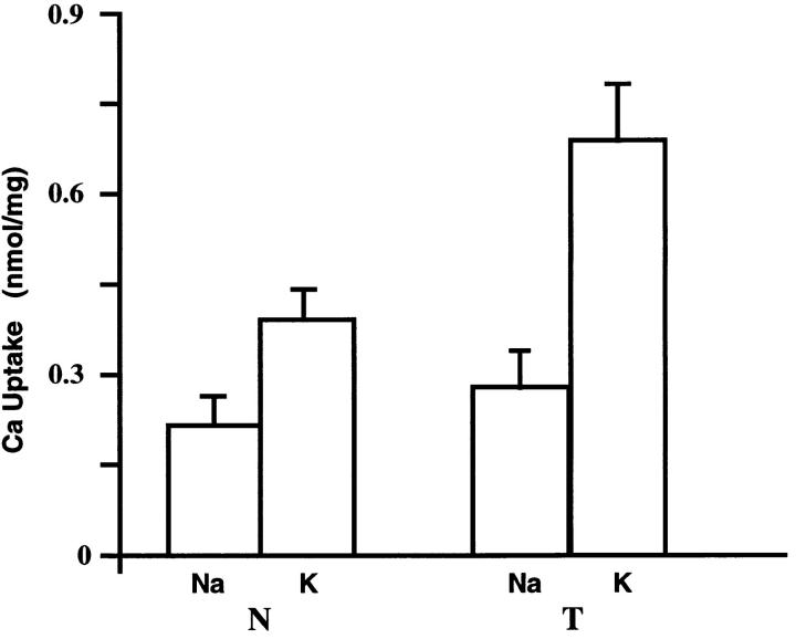
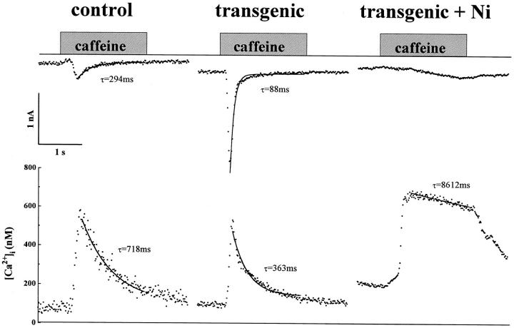
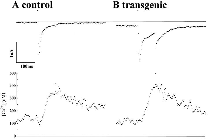
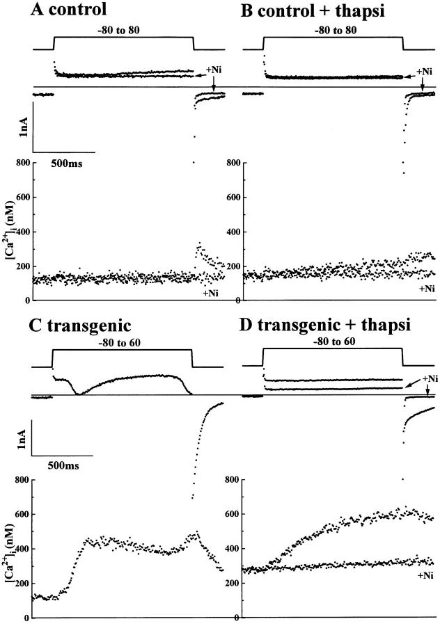
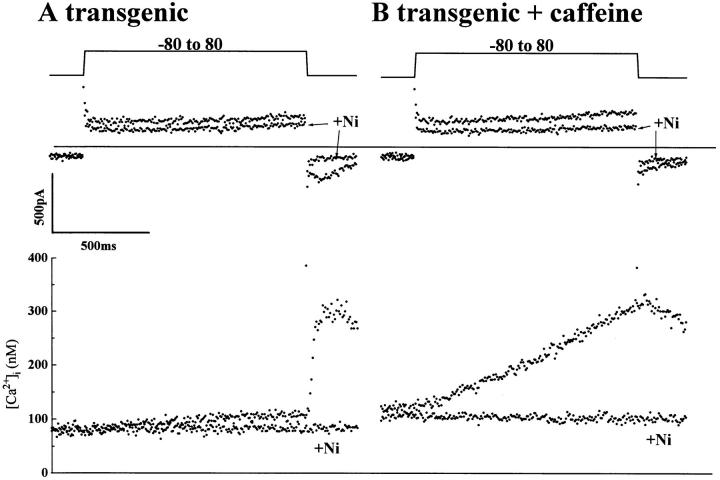
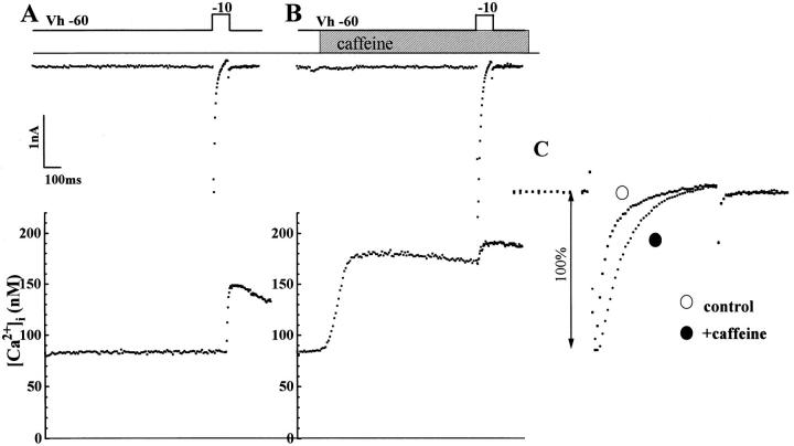
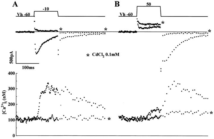
We have produced transgenic mice which overexpress cardiac Na(+)-Ca2+ exchange activity. Overexpression has been assessed by Western blot, Northern blot, and immunofluorescence. Functional overexpression was analyzed using membrane vesicles and isolated ventricular myocytes. In whole cell clamped myocytes dialyzed with 0.1-0.2 mM Fura-2, the magnitude of ICa and Ca2+i-transient triggered by ICa or caffeine were not significantly different in transgenic vs. control myocytes. In transgenic myocytes, activation of ICa, however, was followed by a large slowly inactivating transient inward current representing INa-Ca. This current depended on Ca2+ release as it was abolished when sarcoplasmic reticulum (SR) Ca2+ was depleted using thapsigargin. Cai-transients triggered by rapid application of 5 mM caffeine, even though equivalent in control and transgenic myocytes, activated larger INa-Ca (approximately 5 pA/pF at -90 mV) in transgenic vs. control myocytes (1.5 pA/pF). The decay rate of caffeine-induced Ca2+i-transient and INa-Ca was 2.5 times faster in transgenic than in control myocytes. 5 mM Ni2+ was equally effective in blocking INa-Ca in control or transgenic myocytes. In 9 out of 26 transgenic myocytes, but none of the controls, Ca2+ influx via the exchanger measured at +80 mV caused a slow rise in [Ca2+]i triggering rapid release of Ca2+ from the SR, SR Ca2+ release triggered by the exchanger at such potentials was accompanied by activation of transient current in the inward direction. In 2 mM Fura-2-dialyzed transgenic myocytes caffeine-triggered Cai-transients failed to activate INa-Ca even though the kinetics of inactivation of ICa slowed significantly in caffeine-treated myocytes. In 0.1 mM Fura-2-dialyzed transgenic myocytes 100 microM Cd2+ effectively blocked ICa and suppressed Cai-transients at -10 or +50 mV. Our data suggests that in myocytes overexpressing the exchanger, the content of intracellular Ca2+ pools and the signaling of its release by the Ca2+ channel vis-à-vis the Na(+)-Ca2+ exchanger were not significantly altered despite an up to ninefold increase in the exchanger activity. We conclude that the exchanger remains functionally excluded from the Ca2+ microdomains surrounding the DHP/ryanodine receptor complex.


Figures

Figure 1
Figure 1
Transgene construct. The open reading frame (ORF ) of the Na+-Ca2+ exchanger was under the control of the α-MHC promoter. For details, see text.
Figure 2
Figure 2
Na+-Ca2+ exchanger transcript expression in transgenic mice (A) RNA was isolated from the hearts of transgenic (T ) mice or nontransgenic (N ) littermates from transgenic mouse lines A, C, D, F, H, and G, as indicated. After hybridization, the Northern blot was exposed to film for 1 h. (B ) Northern blot analysis was performed using RNA isolated from the heart (H ), lung (L), brain (B), and skeletal muscle (S) of transgenic mice (T ) or nontransgenic littermates (N ) from line H. After hybridization with an NCX1 cDNA probe, the Northern blot was exposed to film for 6 h. 10 μg of total RNA was run in each lane.
Figure 3
Figure 3
Immunoreactivity of Na+-Ca2+ exchanger protein in transgenic mouse hearts. Shown is Western blot reacted with a polyclonal antibody to the canine cardiac Na+-Ca2+ exchanger. Loaded in each lane was canine cardiac sarcolemmal membranes (10 μg) as a positive control or 50 μg of cardiac homogenate protein from transgenic (T ) mice or nontransgenic (N ) littermates from mouse lines D and A. Antibody dilution was 1/1,000.
Figure 4
Figure 4
Confocal microscope images of isolated mouse myocytes immunolabeled with monoclonal antibodies (R3F1) against the Na+-Ca2+ exchange protein. Both cells were incubated under the same conditions. The gain of the confocal microscope was set to the lowest level to allow the control cell (top) to be just visible. Under these conditions the transgenic myocytes (bottom) have intense labeling of the surface sarcolemma and t-tubules indicating considerable expression of Na+-Ca2+ exchanger protein in the membrane. Also intensely labeled is the area surrounding the nuclei which presumably represents the Golgi. Magnification, 660×.
Figure 5
Figure 5
Na+-Ca2+ exchange activity in isolated membranes. 45Ca2+ uptake into Na+-loaded membrane vesicles was determined in the presence (K+) or absence (Na+) of an outwardly directed Na+ gradient. The external uptake media in these cases contained KCl or NaCl, respectively. The difference in 45Ca2+ uptake from the K+ or Na+ media is taken as Na+-Ca2+exchange activity. Vesicles were prepared from the hearts of transgenic ( T  ) or nontransgenic littermates (N). Media contained 10 μM Ca2+ and the uptake period was 3 s. n = 3 and error bars represent the S.D.
Figure 6
Figure 6
Caffeine-induced Cai-transients and INa-Ca in control and transgenic myocytes. Na+-Ca2+ exchange current was activated by Ca2+-release from the SR triggered by rapid application of 5 mM caffeine at a holding potential of −90 mV. INa-Ca (top) and Ca2+ i -transient (bottom) recorded from a control myocyte. The timing of caffeine application is indicated by the shaded bar. Similar recordings from a transgenic myocyte in the absence and presence of 5 mM NiCl2 as indicated. Cell capacitance of control and transgenic myocytes were 257.9 pF and 239.7 pF, respectively. Fura-2 concentration was 0.2 mM. Data was obtained at room temperature (∼25°C).
Figure 7
Figure 7
ICa-induced Ca2+ release activated “transient inward current” in transgenic but not in control myocytes. Whole cell ICa and Cai-transient were simultaneously recorded from nontransgenic (control, A) and transgenic (B) myocytes. Myocytes were dialyzed with 0.1 mM Fura-2 through patch pipette. ICa were activated by giving depolarizing pulses to −10 mV from a holding potential of −60 mV every 10 s. Cell capacitance in A was 151 pF and in B was 235 pF.
Figure 8
Figure 8
Cai-transients and INa-Ca currents in control and transgenic myocytes in the presence and absence of thapsigargin. The activity of Na+-Ca2+ exchanger activity in the Ca2+ influx mode was measured by applying depolarizing pulses to positive potentials near ECa. (A) Superimposed current traces (top) and the simultaneously measured [Ca2+]i (bottom) during the application of a depolarizing pulse to +80 mV in control myocyte in the presence and absence of 5 mM NiCl2. (B) Similar recordings obtained from the myocyte shown in A after treatment of myocyte with 1.0 μM thapsigargin. (C) Cai-transients and ICa measured at +60 mV in a transgenic myocyte overexpressing Na-Ca exchanger. (D) The superimposed recordings from the same transgenic myocyte as shown in C after treatment with 1.0 μM thapsigargin in the presence and absence of 5 mM NiCl2. Cell capacitance was 151 pF (A and B) and 228 pF (C and D). Myocytes were dialyzed with 0.2 mM Fura-2 concentration.
Figure 9
Figure 9
Enhancement of Ca2+ influx by Na+-Ca2+ exchanger after impairing the Ca2+ storage capacity of the SR by caffeine. (A) Exchanger-dependent currents and rise in [Ca2+]i in transgenic myocyte were elicited by depolarization from −80 to 80 mV. In this myocyte, large depolarization did not cause significant rise in [Ca2+]i, although repolarization from 80 to −80 mV triggered ICa-dependent Ca2+ release, which, in turn, activated INa-Ca inward current. (B) Membrane currents and changes in [Ca2+]i recorded for the same transgenic myocyte (shown in A) in the presence of 5 mM caffeine. The intracellular rise in Ca2+ with depolarization was significantly higher in the presence of caffeine and repolarization no longer triggered Ca2+-release because SR Ca2+ pools were depleted in presence of caffeine. Cell capacitance in A and B was 214 pF. Fura-2 concentration in the patch pipette was 0.2 mM.
Figure 10
Figure 10
Dialysis of transgenic myocytes with high concentrations of Ca2+ buffer does not block cross signaling between Ca2+ channel and ryanodine receptor but blocks activation of exchanger with caffeine-induced Ca2+ release. Ca2+ release by caffeine (5 mM) did not activate inward Na+-Ca2+ exchange current at −90 mV (middle, in a cell dialyzed with 2 mM Fura-2) but altered the rate of inactivation of ICa in panel A (normalized Ca2+ current tracings before and after 600-ms exposure to caffeine are compared in panel C ). Depletion of SR Ca2+ pools by caffeine slowed the inactivation kinetics of ICa even though the rise in [Ca2+]i failed to activate INa-Ca. Cell capacitance was 178.1 pF.
Figure 11
Figure 11
Cd2+ blocks Ca2+ release by blocking ICa at −10 and +50 mV in transgenic myocytes. Membrane currents and Cai-transients activated by depolarizing pulses in transgenic myocytes dialyzed with 0.2 mM Fura-2 were recorded at 10-s intervals before and following addition of 0.1 mM Cd2+(*). ICa, tail current, and Ca2+ i transient activated by test pulses from −60 to −10 mV (A) or to 50 mV (B) were abolished within a few 100 ms by 100 μM Cd2+. Cd2+ increased the net membrane in the outward direction (*) at +50 mV and blocked the Ca2+ transient. The current traces were not leak-subtracted, so as not to suppress the approximately linear components of INa-Ca. Cell capacitance was 228 pF. Fura-2 concentration in dialyzing patch pipette was 0.2 mM.

References

    1. Adachi-Akahane S, Cleemann L, Morad M. Cross-signaling between L-type Ca2+channels and ryanodine receptors in rat ventricular myocytes. J Gen Physiol. 1996;108:435–454. - PMC - PubMed
    1. Callewaert G. Excitation-contraction coupling in mammalian cardiac cells. Cardiovasc Res. 1992;26:923–932. - PubMed
    1. Callewaert G, Cleemann L, Morad M. Caffeine-induced Ca2+-release activates Ca2+ extrusion via Na+-Ca2+exchanger in cardiac myocytes. Am J Physiol. 1989;257:C147–152. - PubMed
    1. Chen F, Mottino G, Klitzner TS, Philipson KD, Frank JS. Distribution of the Na+/Ca2+exchange protein in developing rabbit myocytes. Am J Physiol. 1995;268:C1126–C1132. - PubMed
    1. Chomczynski P, Sacchi N. Single step method of RNA isolation by acid guanidinium thiocyanate-phenol-chloroform extraction. Anal Biochem. 1987;162:156–159. - PubMed

Publication types