. 1997 Jul 22;94(15):7799-806.
doi: 10.1073/pnas.94.15.7799.

Evolution by the birth-and-death process in multigene families of the vertebrate immune system

Affiliations

Evolution by the birth-and-death process in multigene families of the vertebrate immune system

M Nei et al. Proc Natl Acad Sci U S A. .

Abstract

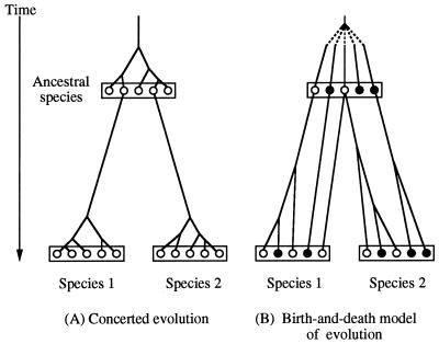
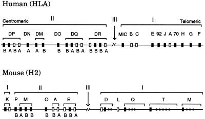
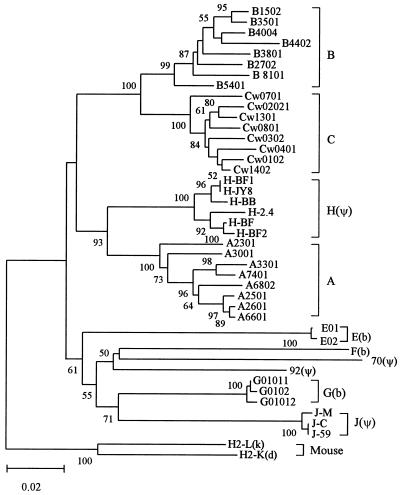
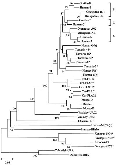
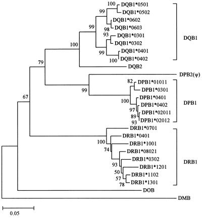
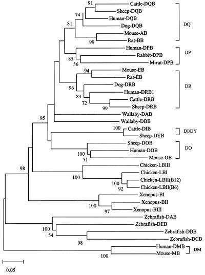
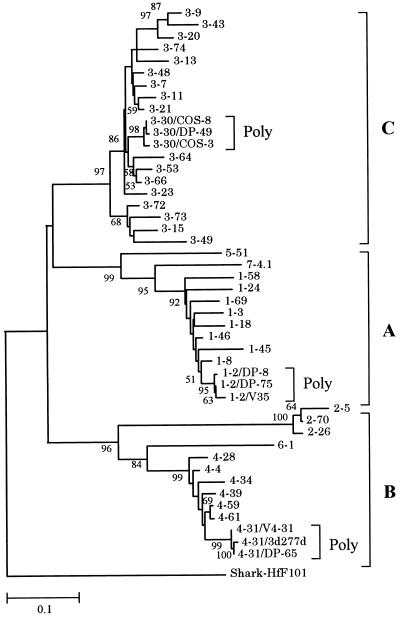
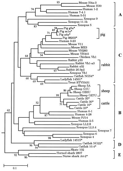
Concerted evolution is often invoked to explain the diversity and evolution of the multigene families of major histocompatibility complex (MHC) genes and immunoglobulin (Ig) genes. However, this hypothesis has been controversial because the member genes of these families from the same species are not necessarily more closely related to one another than to the genes from different species. To resolve this controversy, we conducted phylogenetic analyses of several multigene families of the MHC and Ig systems. The results show that the evolutionary pattern of these families is quite different from that of concerted evolution but is in agreement with the birth-and-death model of evolution in which new genes are created by repeated gene duplication and some duplicate genes are maintained in the genome for a long time but others are deleted or become nonfunctional by deleterious mutations. We found little evidence that interlocus gene conversion plays an important role in the evolution of MHC and Ig multigene families.


Figures

Figure 1
Figure 1
Two different models of evolution of multigene families. ○, Functional gene; •, pseudogene.
Figure 2
Figure 2
Simplified genomic organizations of the human and mouse MHC genes. Only relatively well characterized genes are presented here, and there are many other genes or pseudogenes in both organisms. The recently discovered class Ib gene HLA-HH (21) is located about 4 Mb away from gene F on the telomeric side. (The original authors used the gene symbol HLA-H, but we changed it to HLA-HH to avoid the confusion with an already established Ib locus with the same name.) The number of genes also varies with haplotype in both class I and class II regions. Open boxes refer to polymorphic or classical loci, whereas closed boxes stand for monomorphic or nonclassical loci. Class III genes or other genes unrelated to class I and class II genes are not shown. Class II genes A and B refer to class II α- and β-chain genes, respectively. The MHCs in humans and mice are often called HLA and H2, respectively. The gene maps in this figure are based on information from Trowsdale (22) and other sources.
Figure 3
Figure 3
Phylogenetic tree of human MHC (HLA) class I genes and alleles. The phylogenetic trees presented in this paper are neighbor-joining trees obtained by the computer software mega (35). The tree in this figure was constructed by using Jukes–Cantor distances for the nucleotide sequences (822 bp) of exons 2, 3, and 4, which encode class I MHC domains α1, α2, and α3, respectively. Genes E, F, and G, designated by (b), are functional class Ib loci, whereas genes H, J, 92, and 70, designated by (ψ), are class Ib pseudogenes. Class Ib genes MICA and HH were not included because they are distantly related (see Fig. 4). Two mouse class Ia genes were used as outgroups. The numbers for interior branches refer to the bootstrap values for 500 replications. The bootstrap values less than 50% are not given. The scale at the bottom is in units of nucleotide substitutions per site.
Figure 4
Figure 4
Phylogenetic tree of MHC class I genes from various vertebrate species. This tree was constructed by using p-distances (39) for the amino acid sequences (274 residues) for domains α1, α2, and α3. Class Ib genes are denoted by (b) except for Xenopus genes NC4, NC7, and NC8. Other genes are primarily class Ia genes. Asterisks indicate cDNA data, where the identification of loci or alleles is ambiguous. In this paper we used common species or genus names to designate their MHC genes to make the paper understandable for nonspecialists. The wallaby is a marsupial species; Xenopus is Xenopus laevis.
Figure 5
Figure 5
Phylogenetic tree of human MHC (HLA) class II B genes and alleles. This tree was constructed by using Jukes–Cantor distances for the nucleotide sequences (564 bp) of exons 2 and 3 that encode the class II MHC β-chain domains β1 and β2, respectively. DMB is known to be distantly related to other B genes (Fig. 6).
Figure 6
Figure 6
Phylogenetic tree of MHC class II B genes from various vertebrate species. The tree was constructed by using p-distances for the amino acid sequences (188 residues) of domains β1 and β2. One allele from each locus was used as long as the locus was identifiable. In chicken and Xenopus the class II B loci have not been clearly identified, so that some sequences used may represent different alleles from the same locus. The human and mouse DM B genes, which are known to be distantly related to other class II B genes (31) were used as outgroups. The orthologous genes such as DQ, DP, etc., in different species are bracketed. M-rat, mole rat.
Figure 7
Figure 7
Phylogenetic tree of 40 functional VH genes (loci) and 6 additional polymorphic alleles from humans. This tree was constructed by using Jukes–Cantor distances for the nucleotide sequences (228 bp) of framework regions 1, 2, and 3 (14). Loci 1–2, 3–30, and 4–31 are represented by three alleles. Gene and allelic notations are the same as those of Honjo and Matsuda (53) and the V BASE database (54), from which the sequence data were obtained. The human VH genes are classified into seven families, and the first number of each gene symbol designates the family number, whereas the second number refers to the order of the chromosomal location from the DH gene region.
Figure 8
Figure 8
Phylogenetic tree of 49 VH genes from various vertebrate species. This tree was constructed by using p-distances for the amino acid sequences of framework regions 1, 2, and 3. Most sequence data are from Ota and Nei (14), and additional sequences for cattle, sheep, pigs, and rabbits are from GenBank. The root of the tree was determined by using light chain genes as was done in ref. .

References

    1. Smith G P. Cold Spring Harbor Symp Quant Biol. 1973;38:507–513. - PubMed
    1. Zimmer E A, Martin S L, Beverley S M, Kan Y W, Wilson A C. Proc Natl Acad Sci USA. 1980;77:2158–2162. - PMC - PubMed
    1. Irwin D M, Wilson A C. J Biol Chem. 1990;265:4944–4952. - PubMed
    1. Arnheim N. In: Evolution of Genes and Proteins. Nei M, Koehn R K, editors. Sunderland, MA: Sinauer; 1983. pp. 38–61.
    1. Weiss E H, Mellor A L, Golden L, Fahrner K, Simpson E, Hurst J, Flavell R A. Nature (London) 1983;301:671–674. - PubMed

Publication types