. 1997 Jul 22;94(15):7921-6.
doi: 10.1073/pnas.94.15.7921.

Targeted inactivation of alphai2 or alphai3 disrupts activation of the cardiac muscarinic K+ channel, IK+Ach, in intact cells

Affiliations

Targeted inactivation of alphai2 or alphai3 disrupts activation of the cardiac muscarinic K+ channel, IK+Ach, in intact cells

M O Sowell et al. Proc Natl Acad Sci U S A. .

Abstract

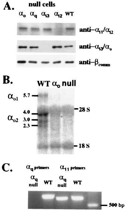
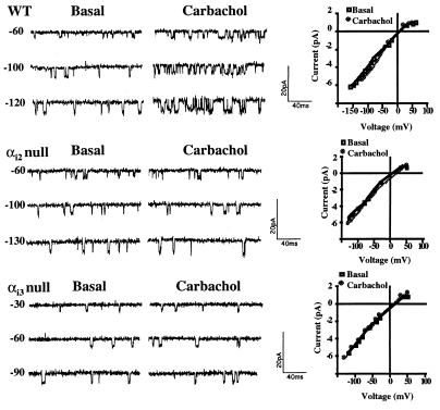
Cardiac muscarinic receptors activate an inwardly rectifying K+ channel, IK+Ach, via pertussis toxin (PT)-sensitive heterotrimeric G proteins (in heart Gi2, Gi3, or Go). We have used embryonic stem cell (ES cell)-derived cardiocytes with targeted inactivations of specific PT-sensitive alpha subunits to determine which G proteins are required for receptor-mediated regulation of IK+Ach in intact cells. The muscarinic agonist carbachol increased IK+Ach activity in ES cell-derived cardiocytes from wild-type cells, in cells lacking alphao, and in cells lacking the PT-insensitive G protein alphaq. In cells with targeted inactivation of alphai2 or alphai3, channel activation by both carbachol and adenosine was blocked. Carbachol-induced channel activation was restored in the alphai2- and alphai3-null cells by reexpressing the previously targeted gene and guanosine 5'-[gamma-thio] triphosphate was able to fully activate IK+Ach in excised membranes patches from these mutants. In contrast, negative chronotropic responses to both carbachol and adenosine were preserved in cells lacking alphai2 or alphai3. Our results show that expression of two specific PT-sensitive alpha subunits (alphai2 and alphai3 but not alphao) is required for normal agonist-dependent activation of IK+Ach and suggest that both alphai2- and alphai3-containing heterotrimeric G proteins may be involved in the signaling process. Also the generation of negative chronotropic responses to muscarinic or adenosine receptor agonists do not require activation of IK+Ach or the expression of alphai2 or alphai3.


Figures

Figure 1
Figure 1
Targeting constructs for inactivation of αi2, αi3, αo, and αq and results of screening by Southern blot analysis. (A) Exon 6 of αi2 was interrupted with a neomycin (neo) resistance cassette. The predicted change in the BamHI (B) restriction digest pattern (increase in size from 4.0 to 8.0 kb) when probed with the indicated gene fragment (shaded bar) is shown below the gene maps. (B Right) The confirmation on Southern blot. For αi3, exon 1 was interrupted with a neo cassette and the ATG initiation codon for translation deleted. Southern blot analysis demonstrated the predicted increase in fragment size (from 5.2 to 6.2 kb) with inactivation of the gene. (C) The targeting construct for inactivation of αo interrupted exon 3 with a neo cassette resulting in a 1.2-kb increase in the BamHI fragment size after homologous recombination. This targeting construct was designed such that both splice variants of αo would be eliminated. (D) For αq, exon 6 was interrupted with a neo cassette resulting in a 1.5-kb increase in the XbaI (X) fragment on Southern blot. Endog, endogenous gene; HR, gene after homologous recombination; SKO, single knockout, heterozygous mutant clone.
Figure 2
Figure 2
Confirmation of α subunit gene inactivations in the mutant ES cell lines. (A) Western blot analysis of membrane protein from undifferentiated WT and α-null cells. (B) Northern blot analysis of total RNA prepared from 7- to 10-day-old differentiated cells and probed with a 1.1-kb NcoI–EcoRI fragment of αo cDNA containing exons 3 through 8 revealed the presence of multiple transcripts in WT cells but none in the αo-null cells. Non-specific binding to 18S and 28S RNA was observed in all lanes. (C) RT-PCR of total RNA was used to assess αq expression and expression of the highly related α11 gene product. An αq product of the predicted size of 655 bp was amplified only in WT cells while the control amplification for α11 detected a product (predicted size of 589 bp) in both WT and the αq-null cells.
Figure 3
Figure 3
Whole cell K+ currents from WT and αi-null ES cell-derived cardiocytes are illustrated. (A) With WT cells a marked increase from basal (a) current was seen after perfusion of the recording chamber with (b) 10 μM carbachol. (B) Addition of the m2 muscarinic receptor antagonist (b) methoctramine (50 nM) did not affect basal current (a) but blocked the subsequent stimulation by carbachol (c). With αi2-null cells (C) and αi3-null cells (D) basal currents (a) were similar to WT cells but did not substantially increase after addition of carbachol (b).
Figure 4
Figure 4
Single channel recordings of the inwardly rectifying K+ channel, IK+Ach, in WT, αi2-null, and αi3-null ES cell-derived cardiocytes. After stable basal recordings were obtained and single channel activities at different voltages recorded, the pipette was perfused with 10 μM of carbachol. Holding voltages (in mV) are shown to the left of the current traces. The I–V relationships for IK+Ach recorded in WT, αi2-null, and αi3-null cells at 150 mM KCl in the absence (basal) and presence of carbachol (10 μM) are similar as shown to the right of the single channel current traces.
Figure 5
Figure 5
Effect of α subunit gene inactivations on receptor-mediated IK+Ach activation. (A) Comparison of IK+Ach activity expressed as NPo before (basal) and after addition of carbachol (10 μM) in WT, α-null cells, and αi-R cells in which the targeted gene has been reexpressed. Bars are means ± SE for 10–20 recordings for WT and αi-null cells and 4 to 5 recordings for αi-R, αo-null, and αq-null cells. (B) Western blot of membrane protein from undifferentiated WT, αi-null cells, and αi-R cells in which the targeted gene (αi2 or αi3) has been reexpressed demonstrate the absence of protein in the null cells but significant protein in the rescued (αi-R) cell lines.
Figure 6
Figure 6
Adenosine’s effect on IK+Ach activation in WT and α-null cell lines. IK+Ach activity was measured before (basal) and after perfusion of the recording pipette with 10 μM adenosine. Bars are means ± SE for 5–8 recordings.
Figure 7
Figure 7
Agonist-independent activation of IK+Ach by 100 μM of GTP[γS] in inside-out patches excised from WT, αi2-null, and αi3-null cells. Shown are typical current traces recorded before (basal) and after addition of GTP[γS] to the bath solution. Activation of IK+ATP was blocked by addition of Mg-ATP (200 μM) to the bath solution. Quantitative estimates of channel activity expressed as NPo are shown in the bottom panel. Bars are the mean ± SE (n = 5–6) before (basal) and after addition of GTP[γS].
Figure 8
Figure 8
The effects of carbachol or adenosine (both 10 μM) on the spontaneous contraction rate of the ES cell-derived cardiocytes were examined in isolated clusters of contracting cells. The effect of pretreatment of cells with PT (0.1 μg/ml for 18 h) on carbachol-induced slowing is also shown. Results are expressed as percent (%) of basal rate calculated as (agonist treated/basal × 100%). Bars are means ± SE for 9–25 paired measurements.

References

    1. Clapham D E. Annu Rev Neurosci. 1994;17:441–464. - PubMed
    1. Kurachi Y. Am J Physiol. 1995;269:C821–C830. - PubMed
    1. Migeon J C, Thomas S L, Nathanson N M. J Biol Chem. 1995;270:16070–16074. - PubMed
    1. Krapivinsky G, Gordon E A, Wickman K, Velimirovic B, Krapivinsky L, Clapham D E. Nature (London) 1995;374:135–141. - PubMed
    1. Wickman K D, Iniguez-Lluhl J A, Davenport P A, Taussig R, Krapivinsky G B, Linder M E, Gilman A G, Clapham D E. Nature (London) 1994;368:255–257. - PubMed

Publication types

MeSH terms

LinkOut - more resources