

Characterization of tumor necrosis factor-deficient mice
- PMID: 9223320
- PMCID: PMC21562
- DOI: 10.1073/pnas.94.15.8093
Characterization of tumor necrosis factor-deficient mice
Abstract
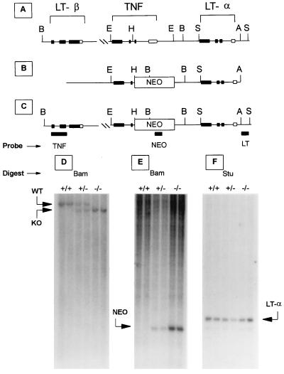
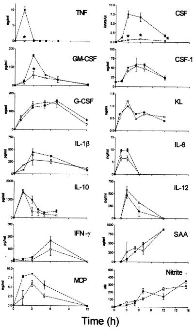
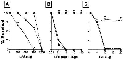
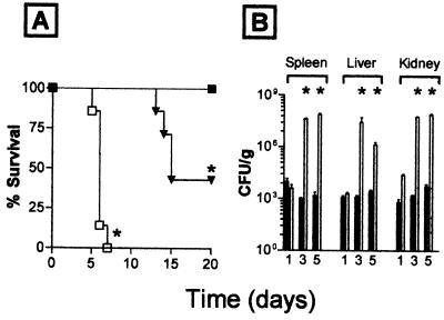
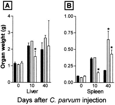
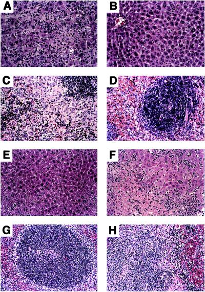
Although tumor necrosis factor (TNF) initially came to prominence because of its anti-tumor activity, most attention is now focused on its proinflammatory actions. TNF appears to play a critical role in both early and late events involved in inflammation, from localizing the noxious agent and amplifying the cellular and mediator responses at the local site and systemically, to editing (e.g., apoptosis) injured cells or effete immune cells and repairing inflammatory damage. We have generated mice deficient in TNF (TNF-/- mice) and have begun to examine the multiple functions attributed to TNF. TNF-/- mice develop normally and have no gross structural or morphological abnormalities. As predicted, they are highly susceptible to challenge with an infectious agent (Candida albicans), are resistant to the lethality of minute doses of lipopolysaccharide (LPS) following D-galactosamine treatment, have a deficiency in granuloma development, and do not form germinal centers after immunization. Phagocytic activity of macrophages appears relatively normal, as do T cell functions, as measured by proliferation, cytokine release, and cytotoxicity. B cell response to thymus-independent antigens is normal, but the Ig response to thymus-dependent antigen is reduced. Surprisingly, cytokine production induced by LPS appears essentially intact, with the exception of reduced colony-stimulating factor activity. Other unexpected findings coming from our initial analysis are as follows. (i) TNF has low toxicity in TNF-/- mice. (ii) TNF-/- mice show an anomalous late response to heat-killed Corynebacterium parvum. In contrast to the prompt response (granuloma formation, hepatosplenomegaly) and subsequent resolution phase in C. parvum-injected TNF+/+ mice, similarly treated TNF-/- mice show little or no initial response, but then develop a vigorous, disorganized inflammatory response leading to death. These results suggest that TNF has an essential homeostatic role in limiting the extent and duration of an inflammatory process-i.e., an anti-inflammatory function. (iii) In contrast to the expectation that TNF+/+ mice and TNF+/- mice would have identical phenotypes, TNF+/- mice showed increased susceptibility to high-dose LPS lethality, increased susceptibility to Candida challenge, and delayed resolution of the C. parvum-induced inflammatory process, indicating a strong gene dose requirement for different actions of TNF.
Figures
References
-
- Beutler B, editor. Tumor Necrosis Factors: The Molecules and Their Emerging Role in Medicine. New York: Raven; 1992.
-
- Aggarwal B B, Vilček J, editors. Tumor Necrosis Factors: Structure, Function, and Mechanism of Action. New York: Dekker; 1992. - PubMed
-
- Beutler B, van Huffel C. Science. 1994;264:667–668. - PubMed
-
- Crowe P D, VanArsdale T L, Walter B N, Ware C F, Hession C, Ehrenfels B, Browning J L, Din W S, Goodwin R G, Smith C A. Science. 1994;264:707–710. - PubMed
MeSH terms
Substances
LinkOut - more resources
Full Text Sources
Other Literature Sources
Molecular Biology Databases
Research Materials
