

Manipulation of T cell costimulatory and inhibitory signals for immunotherapy of prostate cancer
- PMID: 9223321
- PMCID: PMC21563
- DOI: 10.1073/pnas.94.15.8099
Manipulation of T cell costimulatory and inhibitory signals for immunotherapy of prostate cancer
Abstract
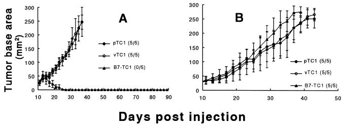
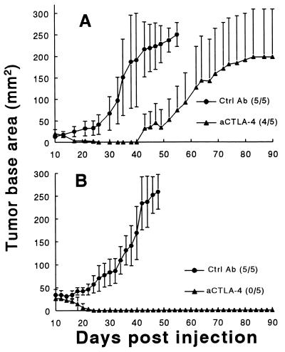
The identification of potentially useful immune-based treatments for prostate cancer has been severely constrained by the scarcity of relevant animal research models for this disease. Moreover, some of the most critical mechanisms involved in complete and proper antitumoral T cell activation have only recently been identified for experimental manipulation, namely, components involved in the costimulatory pathway for T cell activation. Thus, we have established a novel syngeneic murine prostate cancer model that permits us to examine two distinct manipulations intended to elicit an antiprostate cancer response through enhanced T cell costimulation: (i) provision of direct costimulation by prostate cancer cells transduced to express the B7.1 ligand and (ii) in vivo antibody-mediated blockade of the T cell CTLA-4, which prevents T cell down-regulation. In the present study we found that a tumorigenic prostate cancer cell line, TRAMPC1 (pTC1), derived from transgenic mice, is rejected by syngeneic C57BL/6 mice, but not athymic mice, after this cell line is transduced to express the costimulatory ligand B7.1. Also, we demonstrated that in vivo antibody-mediated blockade of CTLA-4 enhances antiprostate cancer immune responses. The response raised by anti-CTLA-4 administration ranges from marked reductions in wild-type pTC1 growth to complete rejection of these cells. Collectively, these experiments suggest that appropriate manipulation of T cell costimulatory and inhibitory signals may provide a fundamental and highly adaptable basis for prostate cancer immunotherapy. Additionally, the syngeneic murine model that we introduce provides a comprehensive system for further testing of immune-based treatments for prostate cancer.
Figures


References
-
- Parker S L, Tong T, Bolden S, Wingo P A. Ca Cancer J Clin. 1997;47:5–27. - PubMed
-
- Keetch D W, Andriole G L. Monogr Urol. 1996;17:31–48.
-
- Sanda M G, Ayyagari S R, Jaffee E M, Epstein J I, Clift S L, Cohen L K, Dranoff G, Pardoll D M, Mulligan R C, Simons J W. J Urol. 1994;151:622–628. - PubMed
-
- Fearon E R, Pardoll D M, Itaya T, Golumbeck P, Levitsky H I, Simons J W, Karasuyama H, Vogelstein B, Frost P. Cell. 1990;60:397–403. - PubMed
-
- Vieweg J, Rosenthal F M, Bannerji R, Heston W D W, Fair W R, Gansbacher B, Gilboa E. Cancer Res. 1994;54:1760–1765. - PubMed
Publication types
MeSH terms
Substances
Grants and funding
LinkOut - more resources
Full Text Sources
Other Literature Sources
Medical
