

Modified cytokeratins expressed on the surface of carcinoma cells undergo endocytosis upon binding of human monoclonal antibody and its recombinant Fab fragment
- PMID: 9223323
- PMCID: PMC21565
- DOI: 10.1073/pnas.94.15.8110
Modified cytokeratins expressed on the surface of carcinoma cells undergo endocytosis upon binding of human monoclonal antibody and its recombinant Fab fragment
Abstract
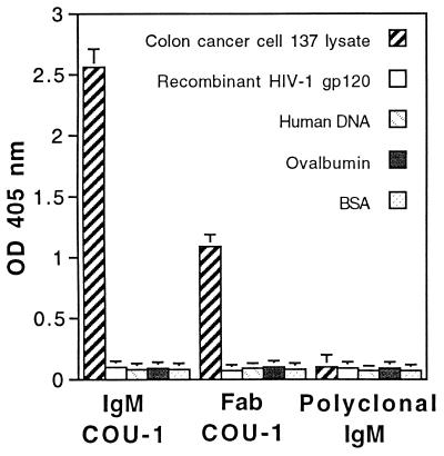
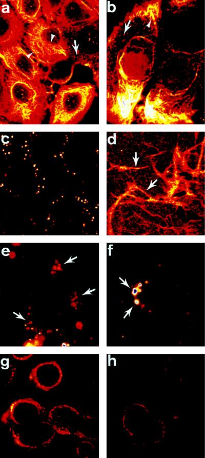
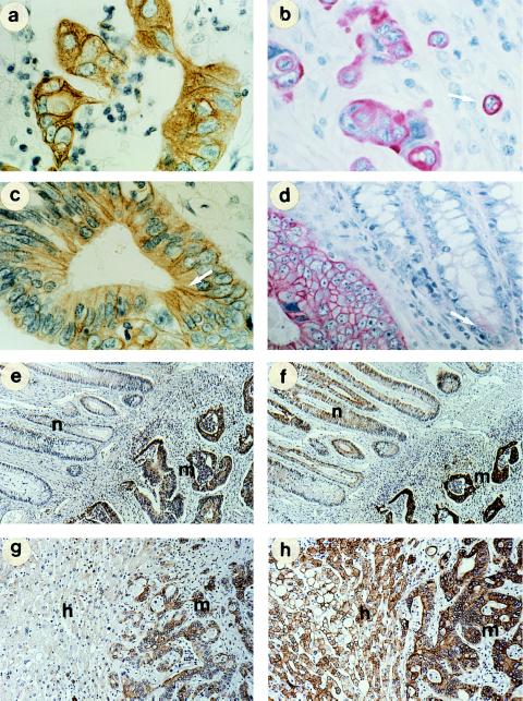
Previously, we have reported on successful imaging of colon, rectal, and pancreatic carcinomas in patients by using a radiolabeled all-human monoclonal antibody, COU-1, directed against modified cytokeratin. To further develop this antibody for use as an immunoconjugate, COU-1 was cloned by phage display selection and the human Fab fragment was expressed in bacteria. Analysis by confocal laser scanning microscopy demonstrated that COU-1 bound in a uniform punctate pattern to the surface of viable carcinoma cells stained at 4 degrees C, and binding increased significantly when cells were cultured on fibronectin, laminin, or collagen IV. In the case of fibronectin, COU-1 staining was particularly enhanced at intercellular junctions. When carcinoma cells were cultured with COU-1 at 37 degrees C for 6 hr, the antibody was found in large perinuclear vesicles and the punctate surface staining was significantly reduced. Similar results were obtained using intact IgM COU-1 and the recombinant Fab fragment. Immunohistological studies indicated that COU-1, in contrast to murine monoclonal antibodies against normal cytokeratin 8 and 18, could differentiate between malignant and normal colon epithelia, and between colon cancer metastasis in the liver and surrounding normal hepatocytes. Within biopsies of malignant tissue, COU-1 exhibited membrane-associated staining of proliferating cells, while resting cells had a filamentous pattern. Thus, modified cytokeratin at the surface of carcinoma cells may represent a new target for immunoconjugates and may explain the promising results of the phase I/II clinical study.
Figures

References
-
- Borup-Christensen P, Erb K, Jensenius J C, Nielsen B, Svehag S-E. Int J Cancer. 1986;37:683–688. - PubMed
-
- Erb K, Borup-Christensen P, Ditzel H, Chemnitz J, Haas H, Jensenius J C. Hybridoma. 1992;11:121–134. - PubMed
-
- Ditzel H, Erb K, Borup-Christensen P, Nielsen B, Jensenius J C. Hum Antibodies Hybridomas. 1991;2:135–141. - PubMed
-
- Borup-Christensen P, Erb K, Ditzel H, Nielsen B, Larsen J K, Svehag S-E, Jensenius J C. AMPIS. 1996;98:674–684. - PubMed
-
- Ditzel H, Rasmussen J W, Erb K, Borup-Christennsen P, Titlestad I, Lassen E, Fenger C, Kronborg O, Jensenius J C. Cancer Res. 1993;53:5920–5928. - PubMed
MeSH terms
Substances
Associated data
- Actions
- Actions
LinkOut - more resources
Full Text Sources
Other Literature Sources
Research Materials
