. 1997 Jul 22;94(15):8162-7.
doi: 10.1073/pnas.94.15.8162.

The hepatitis B virus X gene induces p53-mediated programmed cell death

Affiliations

The hepatitis B virus X gene induces p53-mediated programmed cell death

P Chirillo et al. Proc Natl Acad Sci U S A. .

Abstract

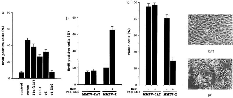
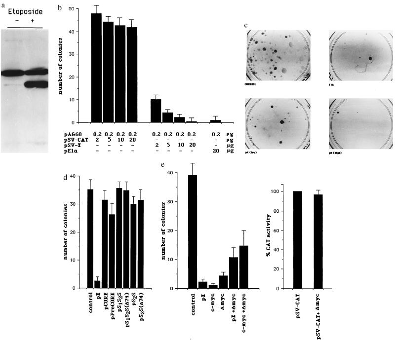
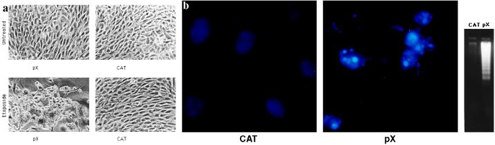
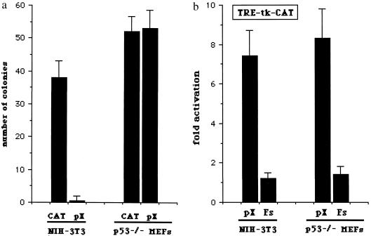
The human hepatitis B virus (HBV) protein pX is a multifunctional regulatory protein that is known to affect both transcription and cell growth. Here we describe induction of apoptosis in NIH 3T3 polyclonal cell lines upon stimulation of pX expression from a dexamethasone inducible mouse mammary tumor virus (MMTV)-X expression vector. The effect of long-term pX expression on the cell survival of mouse fibroblasts was confirmed in colony generation assays. This effect is not shared either by the other HBV products and it is c-myc mediated, as shown by the use of a dominant negative deletion mutant of c-myc. pX also sensitize cells to programmed cell death after exposure to DNA damaging agents. Taking advantage of stable transfectants carrying the p53val135 temperature-sensitive allele, we directly demonstrate that induction of apoptosis by pX requires p53. In p53 null mouse embryo fibroblasts pX activates transcription and confers an evident growth advantage without loss of cell viability. Although pX protein was not detectable in the experimental conditions we used, our results indicate that its expression affects both cell growth and cell death control.


Figures

Figure 1
Figure 1
pX induces both DNA synthesis and loss of cell viability in serum-starved cells. (a) REF-52 cells were transfected with pSV-CAT together with pSV-X, pSV-X(Fs), pCMV-E2F1, and pE1A(12S). The percentage of the CAT positive REF-52 cells also positively displaying BrdUrd was calculated. The data plotted represent the average of three independent experiments. Error bars refer to standard error (SEM). (b) Early passage NIH 3T3 cells were transfected with either the pX expression vector pMMTV-X or the pMMTV-CAT control plasmid. Resistant clones were pooled, made quiescent by serum deprivation, and visualized for BrdUrd incorporation in the presence or absence of dexamethasone. pX transcripts were barely detectable in unstimulated cells and increased several fold after dexamethasone treatment, as assessed by Northern blot analysis (data not shown). Plotted data represent the average of three independent experiments ± standard error (SEM). (c) Serum-starved NIH 3T3 polyclonal populations, obtained as described above, were examined for cell loss by trypan blue dye exclusion and morphological changes at different times after induction of pX or CAT expression. A representative experiment at 72 hr after dexamethasone stimulation is shown (d).
Figure 2
Figure 2
Long-term pX expression reduces cell clonogenic survival in mouse fibroblasts expressing wild-type p53. (a) p53 immunoblotting in untreated and etoposide-treated NIH 3T3 cells using pAb240, which detects both mutant and wild-type p53 in Western blot analysis. The upper band, present in both untreated and etoposide-treated cells, is unspecific. The lower band, whose intensity greatly increases in etoposide-treated cells, is p53. (b) Early passage NIH 3T3 cells were cotransfected with increasing amounts (2–20 μg, as indicated) of either pSV-X or control pSV-CAT expression vectors, pSV-0 as a nonspecific carrier, for a total of 20 μg/ml of DNA, and a fixed amount (200 ng) of the pAG60 geneticin selection plasmid. Resistant colonies were counted after 2–3 weeks. For each experimental condition plotted data represent the average number of colonies obtained in five independent experiments ± standard error (SEM). In c, stained colonies from a representative experiment are shown. (d) Colony generation assays in NIH 3T3 cells using expression vectors for the HBV core, precore/core, large (preS1S2S) and middle (preS2S) envelope proteins (35, 36) and for the truncated envelope proteins with transactivating properties preS1S2S(D74) and preS2S(D74) (35). (e) c-myc mediates pX effects on cell clonogenic survival in NIH 3T3 cells. Cells were cotransfected with pSV-X (10 μg), pCMV-Myc (10 μg), and pCMV-ΔMyc (10 μg), alone or in combination and 200 ng of pAG60 (Left). pCMV-ΔMyc encodes for a c-myc protein carrying a large deletion (amino acids 70–178) in the N-terminal transactivation domain and acts as a dominant negative mutant of wild-type c-myc (37). Colony formation was scored and represented as in a. Cotransfection of pCMV-ΔMyc (2 μg) does not influence the simian virus 40 (SV40) promoter-driven CAT expression (pSV-CAT, 2 μg) (Right).
Figure 3
Figure 3
pX clones are sensitized to p53-mediated apoptosis. (a) Colony regression after exposure to low dosages of the anticancer agent etoposide. pSV-X (2 μg) and pSV-CAT (2 μg) were introduced into NIH 3T3 cells by calcium phosphate coprecipitation and resistant colonies were isolated by selection in G418 as described. Unexpanded pSV-X or pSV-CAT colonies were marked, inspected by microscopy, and photographed 72 hr after treatment with etoposide (Sigma) at the concentration of 0.1 mg/ml. (b) Analysis of chromatin structure (Left and Center) and internucleosomal cleavage of genomic DNA (Right) following treatment with etoposide. Low molecular weight DNA was extracted from equal numbers of cells from either pX or CAT-pooled unexpanded colonies.
Figure 4
Figure 4
p53 status dissects pX properties to induce apoptosis, activate transcription, and alter cell growth. (a) Effect of pX on colony formation in p53-null cells. Early passage p53-deficient MEFs from p53−/− knockout mice were cotransfected with 20 μg of either pSV-X or control pSV-CAT expression vectors, with pSV-0 as a nonspecific carrier, for a total of 20 μg/ml of DNA and 200 ng of pAG60. Colony generation assays were performed, and results are from five independent experiments. (b) Activation of AP1-dependent transcription by pX in serum-starved p53-null cells. p53−/− MEFs were costransfected with 1 μg of the CAT reporter vector pTRE-tk-CAT together with 2 μg of pSV-X, pSV-X(Fs) where appropriate, and pUC18 as nonspecific carrier DNA for a total of 20 μg/ml. The inability of pX to activate transcription from a minimal thymidine kinase (tk) promoter was confirmed using the reporter plasmid pBL2-CATdel.

References

    1. Avantaggiati M L, Natoli G, Balsano C, Chirillo P, Artini M, De Marzio E, Collepardo D, Levrero M. Oncogene. 1993;8:1567–1574. - PubMed
    1. Twu J S, Lai M Y, Chen D S, Robinson W S. Virology. 1993;192:346–350. - PubMed
    1. Balsano C, Avantaggiati M L, Natoli G, De Marzio E, Will H, Perricaudet M, Levrero M. Biochem Biophys Res Commun. 1991;176:985–992. - PubMed
    1. Menzo S, Clementi M, Alfani E, Bagnarelli P, Iacovacci S, Manzin A, Dandri M, Natoli G, Levrero M, Carloni G. Virology. 1993;196:878–882. - PubMed
    1. Doria M, Klein N, Lucito R, Schneider R J. EMBO J. 1995;14:4747–4757. - PMC - PubMed

Publication types

Substances