. 1997 Jul 22;94(15):8208-13.
doi: 10.1073/pnas.94.15.8208.

Interaction between amyloid precursor protein and presenilins in mammalian cells: implications for the pathogenesis of Alzheimer disease

Affiliations

Interaction between amyloid precursor protein and presenilins in mammalian cells: implications for the pathogenesis of Alzheimer disease

W Xia et al. Proc Natl Acad Sci U S A. .

Abstract

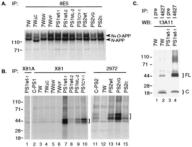
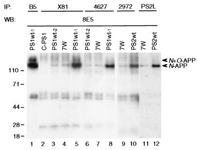
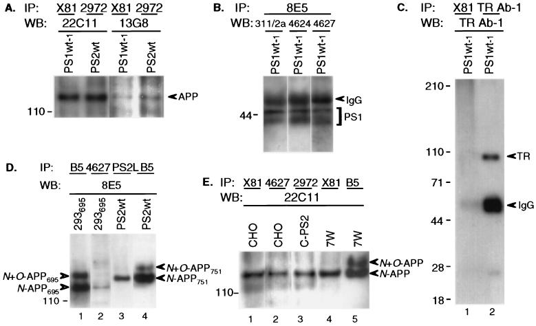
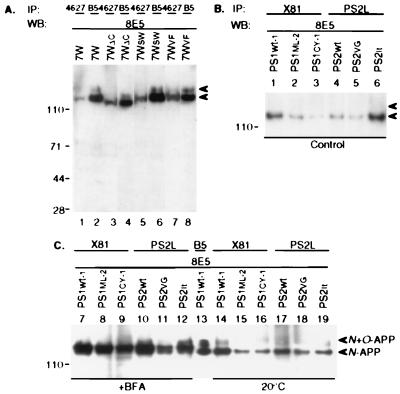
Mutations in the presenilin 1 (PS1) and presenilin 2 (PS2) genes increase the production of the highly amyloidogenic 42-residue form of amyloid beta-protein (Abeta42) in a variety of cell lines and transgenic mice. To elucidate the molecular mechanism of this effect, wild-type (wt) or mutant PS1 and PS2 genes were stably transfected into Chinese hamster ovary cells expressing endogenous or transfected beta-amyloid precursor protein (APP). By immunoprecipitation/Western blot analysis, APP was consistently found to coimmunoprecipitate with PS1 or PS2 proteins. Several distinct PS1, PS2, or APP antibodies precipitated PS-APP complexes that were detectable by blotting with either APP or PS antibodies. Importantly, complex formation could be detected at endogenous protein levels in nontransfected cells. In various Chinese hamster ovary cell lines, the amounts of APP coprecipitated by PS antibodies were proportional to the expression levels of both APP and PS. APP-PS complexes also were recovered from human 293 and HS683 cells. Full maturation of APP was not required for the interaction; most APP molecules complexed with PS were solely N-glycosylated. Treatment of cells with brefeldin A or incubation at 20 degrees C did not block complex formation, suggesting that the association between APP and PS occurs in part in the endoplasmic reticulum. Complex formation was detected for both wt and mutant PS and APP proteins. Deletion of the APP C-terminal domain did not abrogate complex formation, suggesting that the interaction does not occur in the cytoplasmic domains of the proteins. Our results demonstrate that wt and mutant PS1 and PS2 proteins form complexes with APP in living cells, strongly supporting the hypothesis that mutant PS interacts with APP in a way that enhances the intramembranous proteolysis of the latter by a gamma-secretase cleaving at Abeta42.


Figures

Figure 1
Figure 1
Expression of PS and APP proteins in stably transfected CHO cells. Cells were metabolically labeled for 60 min and lysates immunoprecipitated (IP) with the indicated APP (A) or PS1 or PS2 (B) antibodies. (A) APP antibody 8E5 precipitated high levels of APP in all of these APP751-transfected lines. Note the slightly faster migration of both N and N+O-glycosylated APP in cell line 7WΔC, which expresses APP lacking the C-terminal cytoplasmic domain. The slightly lower amounts of APP seen in PS1CY-1 and PS2VG lanes represent gel loading variation and were not consistent. (B) IP with X81 (to PS1) or 2972 (to PS2) precipitated the characteristic PS1 doublet at 43–45 kDa or PS2 doublet at 50–55 kDa (brackets). Both of the PS1 bands have been radiosequenced in the PS1Wt-1 cell line, confirming their identities as SDS-stable conformers of full-length human PS1 (6). The band at ≈44 kDa is a background band that is detected even by X81 preabsorbed with its cognate antigen (lane 1). Endogenous full-length PS1 in 7W, 7WΔC, 7WSW, and 7WVF (lanes 3–6) is not clearly detectable by IP alone. (C) Endogenous hamster PS1 was detected in 7W cell lysate by IP with 4627 followed by Western blotting (WB) with 13A11 (lane 2). Pre, preimmune serum. Transfected human PS1 in line PS1Wt-1 (lane 4) includes full-length (FL) proteins at 43–45 kDa and C-terminal fragments (C) at 18–19 kDa.
Figure 2
Figure 2
Complex formation between PS1 or PS2 and APP in CHO cells. CHO cells stably transfected with PS1 or PS2 were lysed and precipitated (IP) with the indicated PS1 or PS2 antibodies, followed by blotting (WB) with APP antibody 8E5. Lower arrowhead indicates the coprecipitating N-APP polypeptide [compare with the N- and N+O-glycosylated APP precipitated by APP antibody B5 (lane 1)]. IP with antibodies to either PS1 (X81; 4627) or PS2 (2972; PS2L) coprecipitates APP indistinguishably. Bands at 50 and 25 kDa are IgG heavy and light chains.
Figure 3
Figure 3
APP–PS complex formation demonstrated under various conditions. (A) Coprecipitating APP is detectable in PS1Wt-1 or PS2Wt CHO cells using different APP antibodies (22C11; 13G8) for WB. (B) Reverse order of IP/WB: APP antibody (8E5) for IP and various PS1 antibodies (311/2a, 4624, 4627) for WB in PS1Wt-1 cells. IgG bands represent crossreaction of goat anti-rabbit secondary antibody used for WB with primary mouse mAb (8E5) used for IP. (C) IP of PS1Wt-1 CHO cells stably cotransfected with APP, PS1, and human TR using PS1 antibody X81 reveals no coprecipitating TR upon WB with anti-TR (lane 1), whereas both IP and WB with anti-TR reveals TR. Note the expected IgG crossreaction of the anti-rabbit IgG secondary WB antibody with the rabbit primary antibody used for IP. (D) Lane 2, 4627 (PS1) coprecipitates N-glycosylated APP695 (blotted by 8E5) in 293 cells stably transfected with APP695. Lane 3, PS2L (PS2) coprecipitates N-glycosylated APP751 in PS2Wt CHO cells cotransfected with PS2 and APP751. Lanes 1 and 4: IP with B5 as control. (E) Lanes 1 and 2, coprecipitating endogenous APP is detected by 22C11 in nontransfected CHO cells precipitated with X81 or 4627 PS1 antibodies. Lane 3, endogenous APP interacts with transfected PS2 in line C-PS2. Lane 4, transfected APP interacts with endogenous PS1 in line 7W. Lane 5, total APP in 7W cells.
Figure 4
Figure 4
APP–PS complex formation in various cell lines and conditions. (A) IP by 4627 (PS1) or B5 (APP) reveals precipitated APP in CHO single transfectants expressing either wt (7W), C-terminal truncated (7WΔC), Swedish mutant (7WSW), or V698F mutant (7WVF) APP. Note reduced molecular weight of coprecipitated APP in 7WΔC (lanes 3 and 4), as expected. (B) IP by X81 (PS1) or PS2L (PS2) and WB by 8E5 (APP) reveals coprecipitating N-APP (lower arrowhead) in both wt and mutant PS1 and PS2 transfectants. (C) Persistent complex formation in various CHO lines after treatment with BFA (10 μg/ml, l hr) or 20°C temperature block (2.5 hr). The BFA-induced intermediate-sized APP species (27) coprecipitates with PS1 or PS2.

References

    1. Selkoe D J. Science. 1997;275:630–631. - PubMed
    1. Sherrington R, Rogaev E I, Liang Y, Rogaeva E A, Levesque G, et al. Nature (London) 1995;375:754–760. - PubMed
    1. Rogaev E I, Sherrington R, Rogaeva E A, Levesque G, Ikeda M, et al. Nature (London) 1995;376:775–778. - PubMed
    1. Levy-Lahad E, Wasco W, Poorkaj P, Romano D M, Oshima J, Pettingell H, Yu C, Jondro P D, Schmidt S D, Wang K, Crowley A C, Fu Y-H, Guenette S Y, Galas D, Nemens E, Wijsman E M, Bird T D, Schellenberg G D, Tanzi R E. Science. 1995;269:973–977. - PubMed
    1. Borchelt D, Thinakaran G, Eckman C, Lee M, Davenport F, Ratovitsky T, Prada C, Kim G, Seekins S, Yager D, Slunt H, Wang R, Seeger M, Levey A, Gandy S, Copeland N, Jenkins N, Price D, Younkin S, Sisodia S. Neuron. 1996;17:1005–1013. - PubMed

Publication types