. 1997 Jul 28;138(2):225-38.
doi: 10.1083/jcb.138.2.225.

Role of the modular domains of SR proteins in subnuclear localization and alternative splicing specificity

Affiliations

Role of the modular domains of SR proteins in subnuclear localization and alternative splicing specificity

J F Cáceres et al. J Cell Biol. .

Abstract

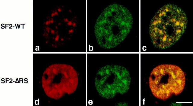
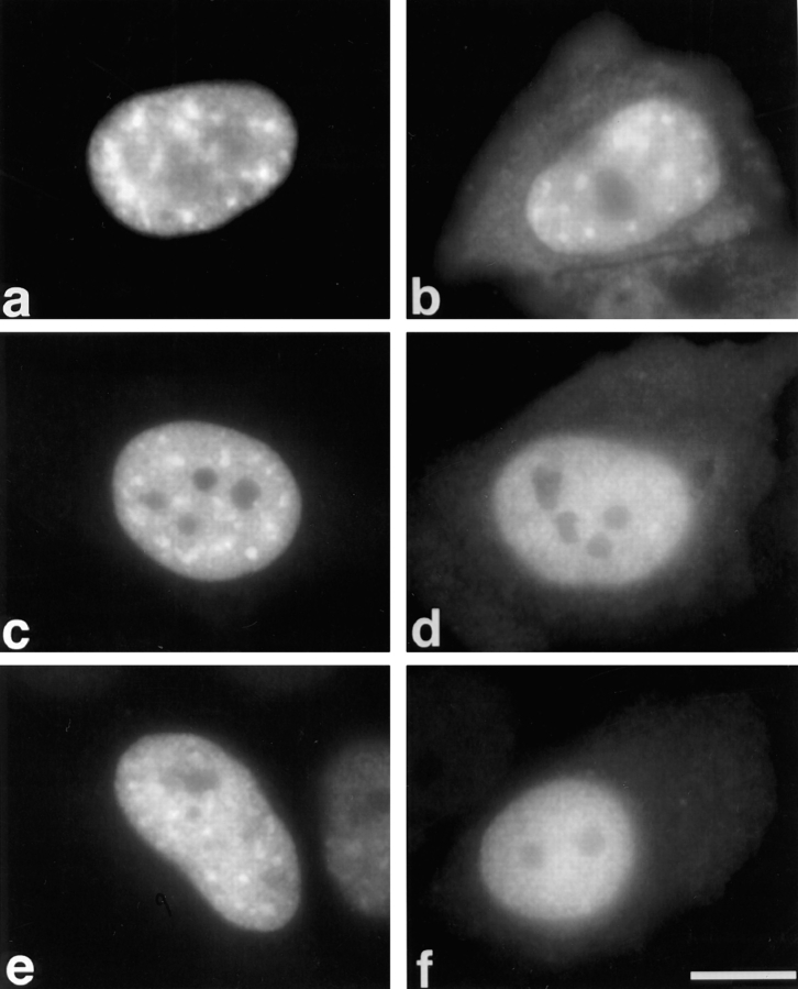
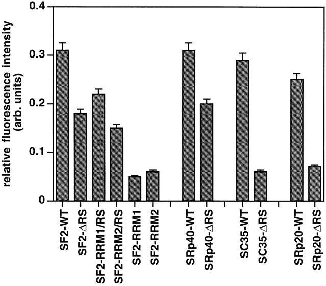
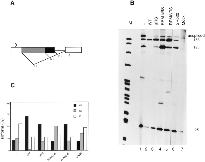
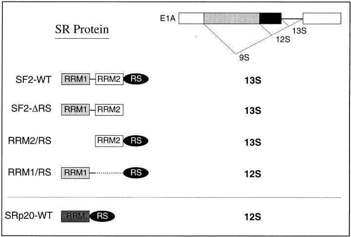
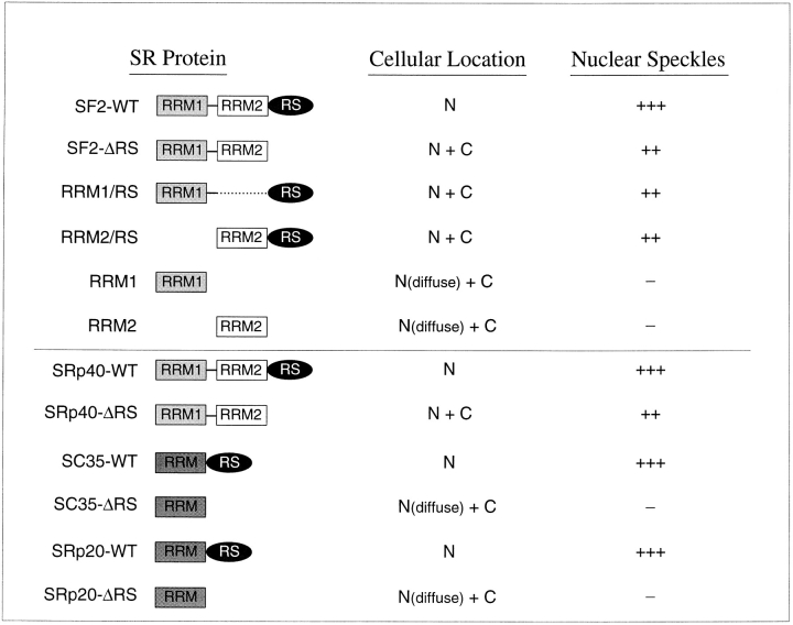
SR proteins are required for constitutive pre-mRNA splicing and also regulate alternative splice site selection in a concentration-dependent manner. They have a modular structure that consists of one or two RNA-recognition motifs (RRMs) and a COOH-terminal arginine/serine-rich domain (RS domain). We have analyzed the role of the individual domains of these closely related proteins in cellular distribution, subnuclear localization, and regulation of alternative splicing in vivo. We observed striking differences in the localization signals present in several human SR proteins. In contrast to earlier studies of RS domains in the Drosophila suppressor-of-white-apricot (SWAP) and Transformer (Tra) alternative splicing factors, we found that the RS domain of SF2/ASF is neither necessary nor sufficient for targeting to the nuclear speckles. Although this RS domain is a nuclear localization signal, subnuclear targeting to the speckles requires at least two of the three constituent domains of SF2/ASF, which contain additive and redundant signals. In contrast, in two SR proteins that have a single RRM (SC35 and SRp20), the RS domain is both necessary and sufficient as a targeting signal to the speckles. We also show that RRM2 of SF2/ASF plays an important role in alternative splicing specificity: deletion of this domain results in a protein that, although active in alternative splicing, has altered specificity in 5' splice site selection. These results demonstrate the modularity of SR proteins and the importance of individual domains for their cellular localization and alternative splicing function in vivo.


Figures

Figure 1
Figure 1
Localization of endogenous SF2/ASF. HeLa cells were fixed, and the cellular localization of SF2/ASF was determined by indirect immunofluorescence with an anti-SF2/ASF monoclonal antibody (Krainer, A.R., unpublished). The protein localizes in a speckled nuclear pattern and is also diffusely distributed throughout the nucleoplasm. (Top) DIC image; (bottom) fluorescence image of the same field. Bar, 10 μm.
Figure 2
Figure 2
Role of SF2/ASF structural domains in cellular localization and distribution in the nuclear speckles. The structure of the SF2/ASF domain-deletion mutants was previously described (Cáceres and Krainer, 1993). (a) SF2-WT, (b) SF2-ΔRS, (c) RRM1/RS, (d) RRM2/RS, (e) RRM1, (f) RRM2. HeLa cells were transfected with plasmids encoding the respective T7-tagged proteins and fixed 24 h after transfection. The localization of the expressed proteins was determined by indirect immunofluorescence with anti-T7 monoclonal antibody and FITC-conjugated secondary antibody. Bar, 5 μm.
Figure 3
Figure 3
Colocalization of transiently expressed SF2/ASF, with or without its RS domain, with endogenous snRNPs. HeLa cells were transiently transfected with plasmids expressing T7-tagged, wild-type SF2/ASF (SF2-WT; top row) or a derivative lacking the RS domain (SF2-ΔRS; bottom row). The cells were fixed 24 h after transfection and analyzed by double-label immunofluorescence using laser scanning confocal microscopy. SF2/ASF or SF2-ΔRS were detected using an anti-T7 mouse monoclonal antibody followed by Texas red–conjugated secondary antibody (a and d). Endogenous snRNPs were detected in the same cell using an anti-Sm human serum followed by FITC-conjugated secondary antibody (b and e). Both SF2-WT and SF2-ΔRS colocalized with endogenous snRNPs in nuclear speckles, as shown by the yellow color when the two signals were superimposed (c and f). Bar, 5 μm.
Figure 4
Figure 4
Cellular localization of other transiently expressed SR proteins with or without their RS domains. HeLa cells were transfected with plasmids encoding the following epitope-tagged proteins: (a) wild-type SRp40, (b) SRp40-ΔRS, (c) wild-type SC35, (d) SC35-ΔRS, (e) wild-type SRp20, and (f) SRp20-ΔRS. The cells were fixed 24 h after transfection and the localization of the expressed proteins was determined as in Fig. 1. Bar, 5 μm.
Figure 5
Figure 5
Quantitative analysis of localization of transfected proteins in nuclear speckles. The relative fluorescence intensity was calculated as described in Materials and Methods. Note that the relative intensity of speckles decreased by ∼30% for SF2/ASF lacking one of its three domains, and by ∼80% when two of the three domains were missing. Results represent average values ± standard deviation from the pooled data of at least two experiments.
Figure 6
Figure 6
Cellular localization of transiently expressed chimeric proteins. HeLa cells were transfected with plasmids encoding the following epitope-tagged proteins: (A) (a) hnRNP A1; (b) A1-RS, a chimeric protein with both RRMs from hnRNP A1 fused to the COOH-terminal RS domain of SF2/ASF. (B) (a) NPc, the nucleoplasmin core domain; (b) NPc-RSSF2, a chimera consisting of NPc fused to the RS domain from SF2/ASF; (c) NPc-RSSRp20, NPc fused to the RS domain of SRp20. The cells were fixed 24 h after transfection and the localization of the expressed proteins was determined as in Fig. 1. (C) Colocalization of transiently expressed NPc-RSSRp20 with endogenous snRNPs. HeLa cells were transiently transfected with plasmid expressing T7-tagged NPc-RSSRp20. The cells were fixed 24 h after transfection and analyzed by double-label immunofluorescence using confocal laser scanning microscopy. NPc-RSSRp20 was detected using an anti-T7 mouse monoclonal antibody followed by Texas red–conjugated secondary antibody (a). Endogenous snRNPs were detected in the same cell using an anti-Sm human serum followed by FITC-conjugated secondary antibody (b). NPc-RSSRp20 colocalized with endogenous snRNPs in nuclear speckles, as shown by the yellow color when the two signals were superimposed (c). Bar, 5 μm.
Figure 6
Figure 6
Cellular localization of transiently expressed chimeric proteins. HeLa cells were transfected with plasmids encoding the following epitope-tagged proteins: (A) (a) hnRNP A1; (b) A1-RS, a chimeric protein with both RRMs from hnRNP A1 fused to the COOH-terminal RS domain of SF2/ASF. (B) (a) NPc, the nucleoplasmin core domain; (b) NPc-RSSF2, a chimera consisting of NPc fused to the RS domain from SF2/ASF; (c) NPc-RSSRp20, NPc fused to the RS domain of SRp20. The cells were fixed 24 h after transfection and the localization of the expressed proteins was determined as in Fig. 1. (C) Colocalization of transiently expressed NPc-RSSRp20 with endogenous snRNPs. HeLa cells were transiently transfected with plasmid expressing T7-tagged NPc-RSSRp20. The cells were fixed 24 h after transfection and analyzed by double-label immunofluorescence using confocal laser scanning microscopy. NPc-RSSRp20 was detected using an anti-T7 mouse monoclonal antibody followed by Texas red–conjugated secondary antibody (a). Endogenous snRNPs were detected in the same cell using an anti-Sm human serum followed by FITC-conjugated secondary antibody (b). NPc-RSSRp20 colocalized with endogenous snRNPs in nuclear speckles, as shown by the yellow color when the two signals were superimposed (c). Bar, 5 μm.
Figure 7
Figure 7
Role of SF2/ASF structural domains in regulating alternative splicing of adenovirus E1A pre-mRNA. (A) Diagram of the E1A reporter gene. The alternative 5′ splice sites and splicing events that generate 13S, 12S, and 9S mRNAs are shown schematically. The location of the exon primers used for RT-PCR analysis is shown. (B) Alternative splicing activity of the SF2/ASF domain-deletion mutants. Each of the indicated SF2/ ASF mutant and control proteins was overexpressed from plasmids cotransfected with the E1A reporter gene. RNA was harvested 24 h after transfection and analyzed by RT-PCR with a labeled forward primer, denaturing PAGE, and autoradiography, as described in Materials and Methods. The positions of the unspliced pre-mRNA, and of 13S, 12S, and 9S spliced mRNAs are indicated on the right. The background products obtained from mock-transfected cells are shown in lane 7. End-labeled DNA size markers are shown in lane M. (C) Quantitation of E1A mRNA isoforms in transfected cells. The relative amounts of 13S, 12S, and 9S E1A mRNAs were calculated from the data in B, using a PhosphorImager, and the percentage of each isoform is shown.
Figure 8
Figure 8
Summary of the role of SR protein domains on E1A alternative splicing specificity in vivo. The domain structures of SF2/ASF and domain-deletion mutants thereof, and that of SRp20, are shown schematically. The predominant E1A isoform generated upon overexpression of each protein is shown on the right. The presence of RRM2 of SF2/ASF correlates with use of the proximal 13S 5′ splice site, whereas proteins that lack this atypical RRM cause preferential use of the 12S 5′ splice site.
Figure 9
Figure 9
Summary of the localization of SR protein variants. The domain structures of the SF2/ASF wild-type and mutant proteins are shown schematically above the line, and those of SRp40, SC35, and SRp20, with or without an RS domain, are shown below the line. The observed cellular location of the expressed proteins is indicated as N (nuclear) or C (cytoplasmic). In all cases, the expressed proteins were excluded from nucleoli. Subnuclear accumulation in speckles is indicated by + or − signs, with the number of + signs reflecting the quantitations shown in Fig. 5. Proteins that did not accumulate in speckles gave a diffuse nuclear pattern.

References

    1. Amrein H, Hedley ML, Maniatis T. The role of specific protein-RNA and protein-protein interactions in positive and negative control of pre-mRNA splicing by Transformer 2. Cell. 1994;76:735–746. - PubMed
    1. Ayane M, Preuss U, Köhler G, Nielsen PJ. A differentially expressed murine RNA encoding a protein with similarities to two types of nucleic acid binding motifs. Nucleic Acids Res. 1991;19:1273–1278. - PMC - PubMed
    1. Bauren G, Wieslander L. Splicing of Balbiani ring 1 gene pre-mRNA occurs simultaneously with transcription. Cell. 1994;76:183–192. - PubMed
    1. Beyer AL, Osheim YN. Splice site selection, rate of splicing, and alternative splicing on nascent transcripts. Genes Dev. 1988;2:754–765. - PubMed
    1. Birney E, Kumar S, Krainer AR. Analysis of the RNA-recognition motif and RS and RGG domains: conservation in metazoan pre-mRNA splicing factors. Nucleic Acids Res. 1993;21:5803–5816. - PMC - PubMed

Publication types