

Clinical and serologic manifestations of autoimmune disease in MRL-lpr/lpr mice lacking nitric oxide synthase type 2
- PMID: 9236188
- PMCID: PMC2199001
- DOI: 10.1084/jem.186.3.365
Clinical and serologic manifestations of autoimmune disease in MRL-lpr/lpr mice lacking nitric oxide synthase type 2
Abstract
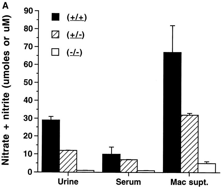
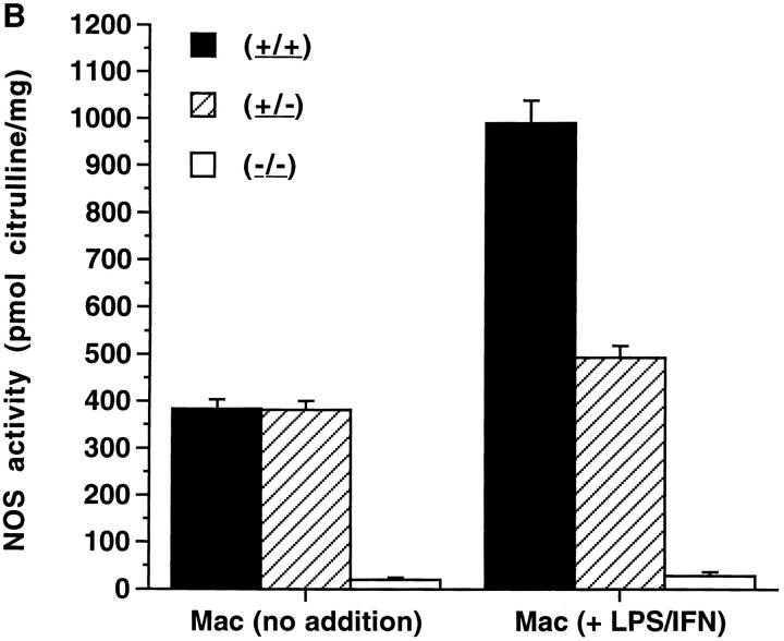
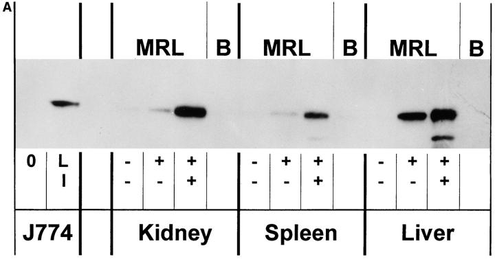
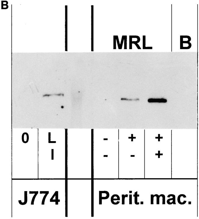
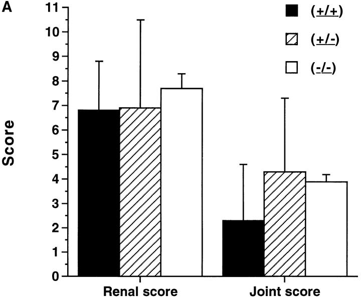
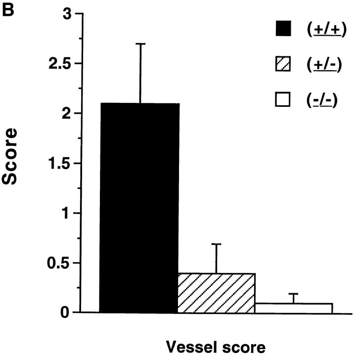
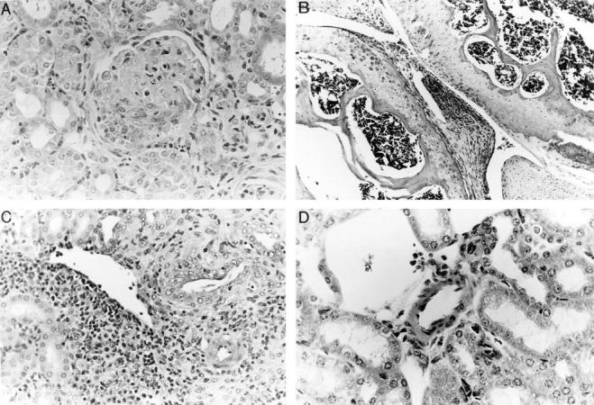
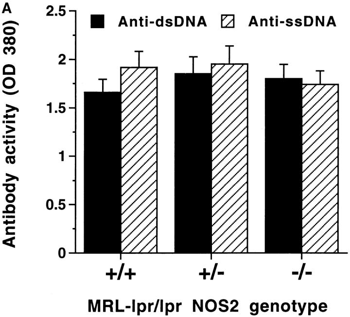
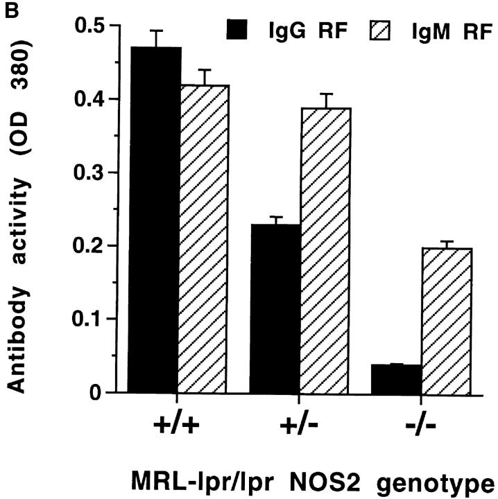
Nitric oxide (NO) is an important mediator of the inflammatory response. MRL-lpr/lpr mice overexpress inducible nitric oxide synthase (NOS2) and overproduce NO in parallel with the development of an autoimmune syndrome with a variety of inflammatory manifestations. In previous studies, we showed that inhibiting NO production with the nonselective nitric oxide synthase (NOS) inhibitor NG-monomethyl-arginine reduced glomerulonephritis, arthritis, and vasculitis in MRL-lpr/lpr mice. To define further the role of NO and NOS2 in disease in MRL-lpr/lpr mice, mice with targeted disruption of NOS2 were produced by homologous recombination and bred to MRL-lpr/lpr mice to the N4 generation. MRL-lpr/lpr littermates homozygous for disrupted NOS2 (-/-), heterozygous for disrupted NOS2 (+/-), or wildtype (+/+) were derived for this study. Measures of NO production were markedly decreased in the MRL-lpr/lpr (-/-) mice compared with MRL-lpr/lpr (+/+) mice, with intermediate production by the MRL-lpr/lpr (+/-) mice. There was no detectable NOS2 protein by immunoblot analysis of the spleen, liver, kidney, and peritoneal macrophages of the (-/-) animals, whereas that of (+/+) was high and (+/-) intermediate. The (-/-) mice developed glomerular and synovial pathology similar to that of the (+/-) and (+/+) mice. However, (-/-) mice and (+/-) mice had significantly less vasculitis of medium-sized renal vessels than (+/+) mice. IgG rheumatoid factor levels were significantly lower in the (-/-) mice as compared with (+/+) mice, but levels of anti-DNA antibodies were comparable in all groups. Our findings show that NO derived from NOS2 has a variable impact on disease manifestations in MRL-lpr/lpr mice, suggesting heterogeneity in disease mechanisms.
Figures
Similar articles
-
Nitric oxide as an inflammatory mediator in autoimmune MRL-lpr/lpr mice.Environ Health Perspect. 1998 Oct;106 Suppl 5(Suppl 5):1131-7. doi: 10.1289/ehp.98106s51131. Environ Health Perspect. 1998. PMID: 9788887 Free PMC article. Review.
-
The role of nitric oxide in the pathogenesis of spontaneous murine autoimmune disease: increased nitric oxide production and nitric oxide synthase expression in MRL-lpr/lpr mice, and reduction of spontaneous glomerulonephritis and arthritis by orally administered NG-monomethyl-L-arginine.J Exp Med. 1994 Feb 1;179(2):651-60. doi: 10.1084/jem.179.2.651. J Exp Med. 1994. PMID: 7507509 Free PMC article.
-
Peroxynitrite formation and decreased catalase activity in autoimmune MRL-lpr/lpr mice.Mol Med. 2000 Sep;6(9):779-92. Mol Med. 2000. PMID: 11071272 Free PMC article.
-
Modulation of renal disease in MRL/lpr mice by pharmacologic inhibition of inducible nitric oxide synthase.Kidney Int. 2002 Mar;61(3):839-46. doi: 10.1046/j.1523-1755.2002.00230.x. Kidney Int. 2002. PMID: 11849435
-
Genetic basis of autoimmune disease in MRL/lpr mice: dissection of the complex pathological manifestations and their susceptibility loci.Rev Immunogenet. 2000;2(1):154-64. Rev Immunogenet. 2000. PMID: 11324688 Review.
Cited by
-
Inducible nitric oxide synthase is expressed in joints of goats in the late stage of infection with caprine arthritis encephalitis virus.Clin Exp Immunol. 1999 Jul;117(1):70-5. doi: 10.1046/j.1365-2249.1999.00932.x. Clin Exp Immunol. 1999. PMID: 10403918 Free PMC article.
-
Serum nitric oxide (NO) levels in systemic sclerosis patients: correlation between NO levels and clinical features.Clin Exp Immunol. 2003 Dec;134(3):538-44. doi: 10.1111/j.1365-2249.2003.02320.x. Clin Exp Immunol. 2003. PMID: 14632763 Free PMC article.
-
Systems biology of lupus: mapping the impact of genomic and environmental factors on gene expression signatures, cellular signaling, metabolic pathways, hormonal and cytokine imbalance, and selecting targets for treatment.Autoimmunity. 2010 Feb;43(1):32-47. doi: 10.3109/08916930903374774. Autoimmunity. 2010. PMID: 20001421 Free PMC article. Review.
-
The role of nitric oxide in abnormal T cell signal transduction in systemic lupus erythematosus.Clin Immunol. 2006 Feb-Mar;118(2-3):145-51. doi: 10.1016/j.clim.2005.10.016. Epub 2006 Jan 10. Clin Immunol. 2006. PMID: 16406340 Free PMC article. Review.
-
Curcumin, the major component of food flavour turmeric, reduces mucosal injury in trinitrobenzene sulphonic acid-induced colitis.Br J Pharmacol. 2003 May;139(2):209-18. doi: 10.1038/sj.bjp.0705241. Br J Pharmacol. 2003. PMID: 12770926 Free PMC article.
References
-
- Cohen PL, Eisenberg RA. Lpr and gld: single gene models of systemic autoimmunity and lymphoproliferative disease. Annu Rev Immunol. 1991;9:243–269. - PubMed
Publication types
MeSH terms
Substances
Grants and funding
LinkOut - more resources
Full Text Sources
Medical
Molecular Biology Databases
