. 1997 Aug 15;17(16):6094-104.
doi: 10.1523/JNEUROSCI.17-16-06094.1997.

Detection of functional nicotinic receptors blocked by alpha-bungarotoxin on PC12 cells and dependence of their expression on post-translational events

Affiliations

Detection of functional nicotinic receptors blocked by alpha-bungarotoxin on PC12 cells and dependence of their expression on post-translational events

E M Blumenthal et al. J Neurosci. .

Abstract

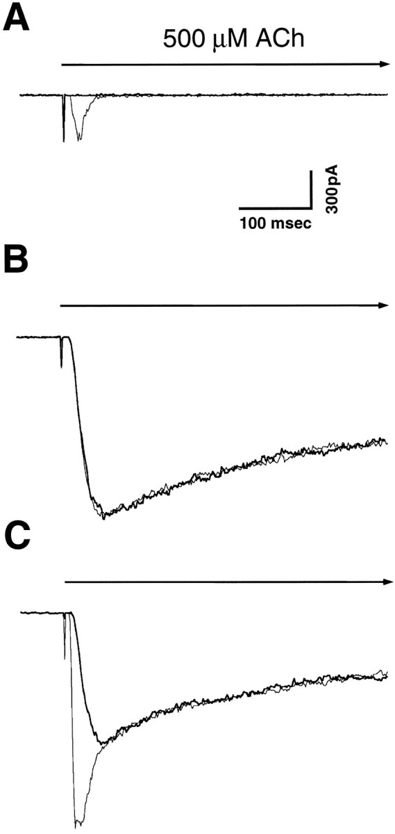
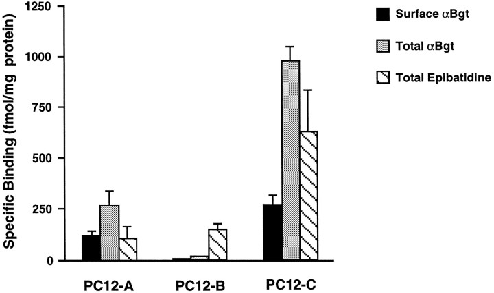
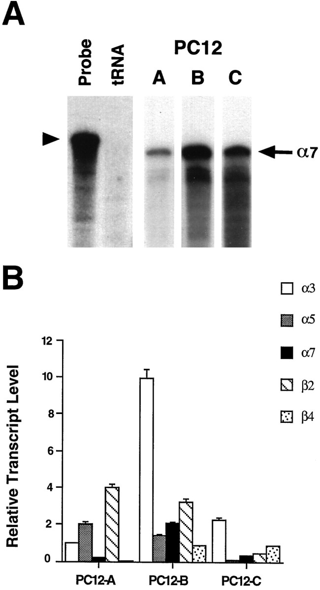
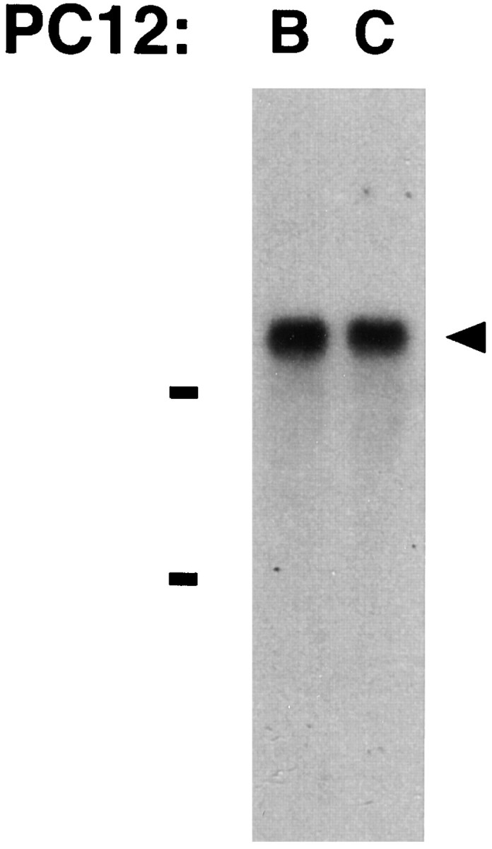
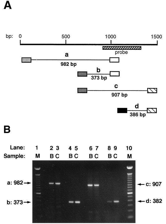
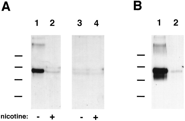
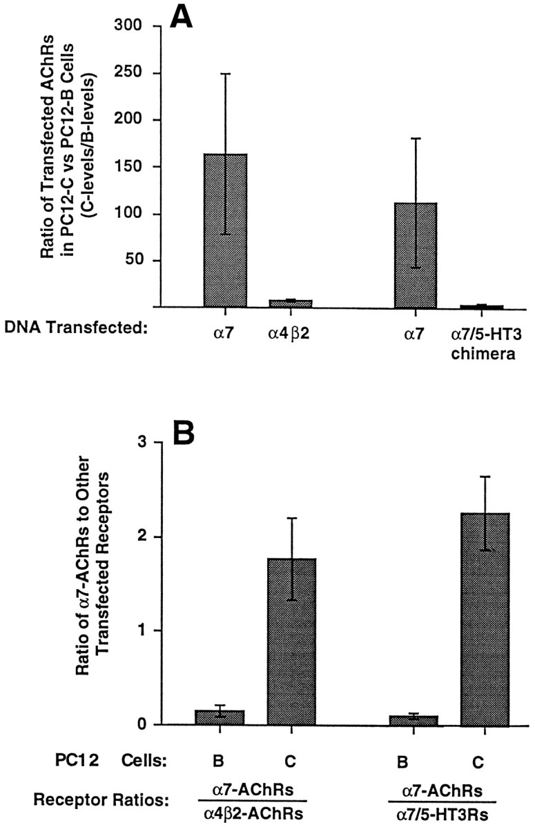
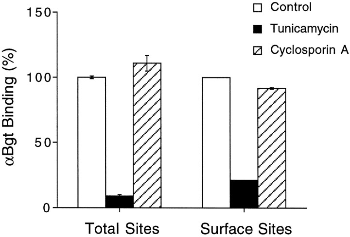
A major class of nicotinic receptors in the nervous system is one that binds alpha-bungarotoxin and contains the alpha7 gene product. PC12 cells, frequently used to study nicotinic receptors, express the alpha7 gene and have binding sites for the toxin, but previous attempts to elicit currents from the putative receptors have failed. Using whole-cell patch-clamp recording techniques and rapid application of agonist, we find a rapidly desensitizing acetylcholine-induced current in the cells that can be blocked by alpha-bungarotoxin. The current amplitude varies dramatically among three populations of PC12 cells but correlates well with the number of toxin-binding receptors. In contrast, the current shows no correlation with alpha7 transcript; cells with high levels of alpha7 mRNA can be negative for toxin binding and yet have other functional nicotinic receptors. Northern blot analysis and reverse transcription-PCR reveal no defects in alpha7 RNA from the negative cells, and immunoblot analysis demonstrates that they contain full-length alpha7 protein, although at reduced levels. Affinity purification of toxin-binding receptors from cells expressing them confirms that the receptors contain alpha7 protein. Transfection experiments demonstrate that PC12 cells lacking native toxin-binding receptors are deficient at producing receptors from alpha7 gene constructs, although the same cells can produce receptors from other transfected gene constructs. The results indicate that nicotinic receptors that bind alpha-bungarotoxin and contain alpha7 subunits require additional gene products to facilitate assembly and stabilization of the receptors. PC12 cells offer a model system for identifying those gene products.


Figures

Fig. 1.
Fig. 1.
αBgt blockade of ACh-evoked currents in PC12 cells. Whole-cell patch clamp was used to measure currents evoked in PC12-A (A), PC12-B (B), and PC12-C (C) cells by rapid application of 500 μm ACh in the presence and absence of αBgt. Each panel shows typical responses from a control cell (light trace) and a cell incubated with 60 nm αBgt for 1–3 hr (heavy trace) superimposed. PC12-A cells display a small, rapidly activating current that quickly desensitizes and is completely blocked by αBgt; PC12-B cells show a large, slowly activating current that is completely insensitive to αBgt; PC12-C cells have both kinds of responses. Holding potential, −60 mV. Calibration bars: horizontal, 100 msec; vertical, 300 pA. The initiation and duration of ACh application are indicated by thevertical deflection and heavy horizontal arrow, respectively.
Fig. 2.
Fig. 2.
Differences among PC12 populations in their relative levels of AChRs that bind αBgt and epibatidine.125I-αBgt was used to measure binding sites on intact cells, whereas both 125I-αBgt and3H-epibatidine were used to quantify sites in cell extracts. The results were normalized for total protein. Nonspecific binding was determined by adding 1 μm αBgt (αBgt assays), 1 μm epibatidine (epibatidine assay), or 250 μm nicotine (both assays) to the binding reactions and was subtracted from the values shown. PC12-A and -C cells express significant amounts of αBgt binding sites both on the surface (solid bars) and in cell extracts (stippled bars), whereas PC12-B cells have few if any specific αBgt binding sites. All three populations have significant epibatidine binding (hatched bars). Data represent the mean ± SEM of triplicate determinations from four to seven independent experiments.
Fig. 3.
Fig. 3.
Relative amounts of AChR gene transcripts in three PC12 populations. RNase protection assays were used to measure the levels of α3, α5, α7, β2, and β4 transcripts in PC12-A–PC12-C cells. A, Representative RNase protection autoradiogram in which 20 μg samples of total RNA were hybridized with a 32P-labeled α7 riboprobe (Probe lane, arrowhead). tRNA (15 μg) served as a negative control; neither it nor probe alone protected a radiolabeled species (tRNA lane). Each PC12 RNA sample protected an α7 band of the expected size (arrow); the levels of α7 mRNA varied among the three cell populations, with the PC12-B cells expressing the most. B, The protected species obtained with each of the five probes was quantified on a Bio-Rad Molecular Imager. The results represent the mean ± SEM for three to seven determinations from a total of three or four RNA preparations and are normalized to the signal obtained for α3 transcript in PC12-A cells.
Fig. 4.
Fig. 4.
Northern blot analysis demonstrating α7 transcripts of equivalent size in PC12-B and PC12-C cells. Total RNA was isolated from each population and subjected to electrophoresis on a formaldehyde–agarose gel followed by capillary blotting onto a nylon membrane. The blot was hybridized with random-primed,32P-labeled probe directed to the cytoplasmic domain of α7. Bars indicate the 28 and 18 S ribosomal RNAs. Both populations express a single α7 transcript ∼6.4 kb in length (arrowhead). The results are qualitative with respect to band intensity and should not be used to assess relative abundance of the transcripts. Similar results were obtained in four other experiments.
Fig. 5.
Fig. 5.
RT-PCR analysis comparing the coding regions of α7 mRNA from PC12-B and -C cells. A, Schematic showing the α7 transcript nucleotide sequence (heavy line) with the start coinciding with the A of the AUG start codon (Seguela et al., 1993). The expected PCR fragments (thin lines) werea (982 bp), b (373 bp), c(907 bp), and d (386 bp). Primer locations corresponded to nucleotide positions 18–38 (lightly stippled box), 627–646 (heavily stippled box), 981–1000 (open box), 1148–1167 (solid box), and 1516–1534 (hatched box). The sequence encoded by the RNase protection probe is indicated by the hatched bar.B, Agarose gels showing the amplified PCR fragments. First-strand cDNA was synthesized using reverse transcriptase and total RNA isolated from PC12-B and -C cells as templates. PCR was performed with the indicated sets of primers. Aliquots of the PCR reactions were run on 1.1% agarose gels, which were stained with ethidium bromide and photographed using a Polaroid camera. Lanes 1 and10, M, Molecular weight standards (100 bp ladder from Gibco-BRL); lanes 2 and 3, PCR fragment a; lanes 4 and 5, fragment b;lanes 6 and 7, fragment c; lanes 8 and 9, fragment d. B, PC12-B RNA as template; C, PC12-C RNA as template. No differences were detected in the fragments amplified from the PC12-B and -C RNA, and in each case the fragment had the full size expected. Similar results were obtained when PCR was performed on cDNA synthesized from two independent RNA preparations.
Fig. 6.
Fig. 6.
Immunoblot analysis showing α7 protein in PC12 AChRs. A, AChRs that bind αBgt were affinity-purified from PC12-B and -C cell extracts by adsorption to αBgt coupled to Actigel beads. The adsorbed material was eluted and analyzed by immunoblots probed with the anti-α7 mAb A7–1 and visualized using a horseradish peroxidase-coupled secondary antibody followed by enhanced chemiluminescence. A single species of about 60 kDa was obtained from PC12-C samples (lane 1). Nicotine at 250 μm (lane 2) blocked adsorption of the component to the αBgt-Actigel, indicating the specificity of the affinity purification. No components were obtained specifically when PC12-B cell extracts were analyzed by similar methods (lanes 3 and 4). Molecular weight standards (Bio-Rad) are as follows: phosphorylase B, 97 kDa; serum albumin, 66 kDa; ovalbumin, 45 kDa; and carbonic anhydrase, 31 kDa. Similar results were obtained in a second experiment using mAb A7–1 as probe and in two experiments using mAb 319 as probe. B, Total α7 protein from PC12-B and -C cell extracts was analyzed by immunoprecipitating protein with the α7-specific mAb 319 coupled to Actigel and probing immunoblots of the purified material with mAb A7–1. A species of 60 kDa component was obtained from both PC12-C (lane 1) and PC12-B (lane 2) cell extracts, although the latter contained much less of the component. Similar results were obtained in a second experiment. The band was absent when rat IgG-Actigel was substituted for the mAb 319-Actigel as a negative control (data not shown).
Fig. 7.
Fig. 7.
Expression of transfected receptor gene constructs in PC12 cells. PC12-B and -C cells were transfected with chicken AChR cDNA constructs encoding either the α7 or the α4 and β2 subunits or, in separate experiments, containing either the α7 or the α7/5-HT3 chimeric receptor subunit. Cell extracts were prepared 20–48 hr later and assayed by RIA for 125I-αBgt binding to receptors tethered either by anti-α7 mAb 318, which recognizes chick but not rat α7 protein, or by anti-myc mAb 9E10, which recognizes the epitope-tagged α7/5-HT3 receptor protein and by RIA for 3H-epibatidine binding to receptors tethered with anti-α4 mAb 289, which recognizes chick but not rat α4 protein. The selectivities of the antibodies required that the receptors contain one or more transfected gene products to be assayed (receptors containing only endogenous gene components would not have been retained). A, Ratio of receptor levels in PC12-C cell extracts divided by that in PC12-B cell extracts obtained from the same experiment. Values represent the mean ± SEM of three or four experiments. Absolute levels of αBgt binding to chick AChRs with α7 subunits in transfected PC12-B and PC12-C cells averaged 6 and 498 fmol/culture, respectively, for the experiments with α4β2 and 4 and 258 fmol/culture, respectively, for the experiments with α7/5-HT3. Levels of epibatidine binding for PC12-B and PC12-C cells were 43 and 288 fmol/culture, respectively, in the α4β2 experiments; levels of αBgt binding to 5-HT3chimeras were 37 and 111 fmol/culture, respectively. PC12-B cells have a large deficiency in the expression of AChRs with α7 subunits compared with PC12-C cells. B, Relative number of αBgt-binding AChRs produced by transfected PC12-B (B) or PC12-C (C) cells with chick α7 subunits (α7-AChRs) divided by either the number of receptors with α4 and β2 subunits (α4β2-AChRs) or α7/5-HT3 chimeric receptor subunits (α7/5-HT3Rs). PC12-B cells are significantly more impaired at expressing αBgt-binding AChRs from the α7 construct than they are at expressing either AChRs from the α4 and β2 constructs (p < 0.03 by Student’s unpaired t test) or receptors from the α7/5-HT3 chimeric construct (p< 0.002).
Fig. 8.
Fig. 8.
Lack of cyclosporin A effects on levels of PC12 receptors binding αBgt. PC12-C cells in culture were treated either with vehicle (Control), tunicamycin (1 μg/ml), or cyclosporin A (10 μm) for 24 hr and then assayed either for αBgt binding in RIAs with mAb 318 (which recognizes the chick but not rat α7 gene product) to immunotether receptors (Total Sites) or for αBgt-binding to intact cells in culture (Surface Sites). Values represent the mean ± SEM of three or four separate experiments and are expressed as percentages of those obtained from control cells. Tunicamycin treatment caused a large reduction in αBgt binding, indicating that receptors both on the cell surface and inside were normally turning over. Cyclosporin A treatment had little effect either on the total number of αBgt-binding receptors assayed or on those present on the cell surface.

Similar articles

Cited by

References

    1. Alkondon M, Albuquerque EX. Diversity of nicotinic acetylcholine receptors in rat hippocampal neurons. I. Pharmacological and functional evidence for distinct structural subtypes. J Pharmacol Exp Ther. 1993;265:1455–1473. - PubMed
    1. Alkondon M, Reinhardt S, Lobron C, Hermsen B, Maelicke A, Albuquerque EX. Diversity of nicotinic acetylcholine receptors in rat hippocampal neurons. II. The rundown and inward rectification of agonist-elicited whole-cell currents and identification of receptor subunits by in situ hybridization. J Pharmacol Exp Ther. 1994;271:494–506. - PubMed
    1. Amy CM, Bennett EL. Increased sodium ion conductance through nicotinic acetylcholine receptor channels in PC12 cells exposed to nerve growth factors. J Neurosci. 1983;3:1547–1553. - PMC - PubMed
    1. Anand R, Peng X, Lindstrom J. Homomeric and native α7 acetylcholine receptors exhibit remarkably similar but non-identical pharmacological properties, suggesting that the native receptor is a heteromeric protein complex. FEBS Lett. 1993;327:241–246. - PubMed
    1. Bertrand D, Galzi JL, Devillers-Thiery A, Bertrand S, Changeux JP. Mutations at two distinct sites within the channel domain M2 alter calcium permeability of neuronal α7 nicotinic receptor. Proc Natl Acad Sci USA. 1993;90:6971–6975. - PMC - PubMed

Publication types

MeSH terms

LinkOut - more resources