. 1997 Aug 15;17(16):6142-51.
doi: 10.1523/JNEUROSCI.17-16-06142.1997.

Increased neuronal endocytosis and protease delivery to early endosomes in sporadic Alzheimer's disease: neuropathologic evidence for a mechanism of increased beta-amyloidogenesis

Affiliations

Increased neuronal endocytosis and protease delivery to early endosomes in sporadic Alzheimer's disease: neuropathologic evidence for a mechanism of increased beta-amyloidogenesis

A M Cataldo et al. J Neurosci. .

Abstract

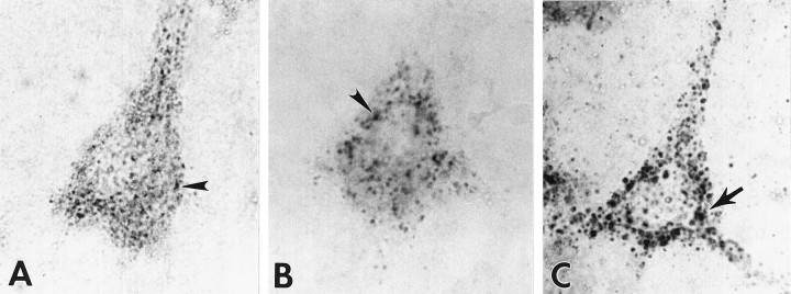
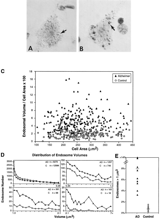
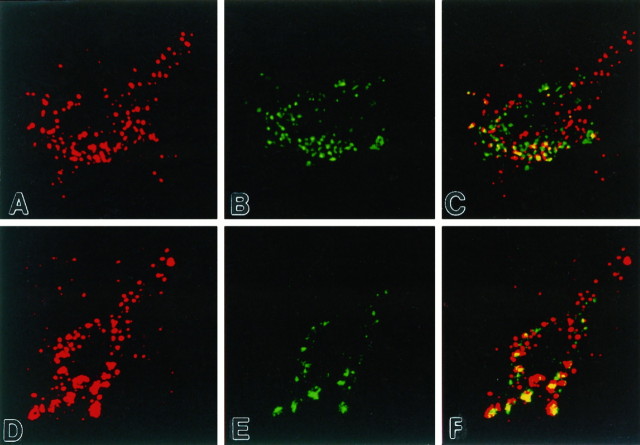
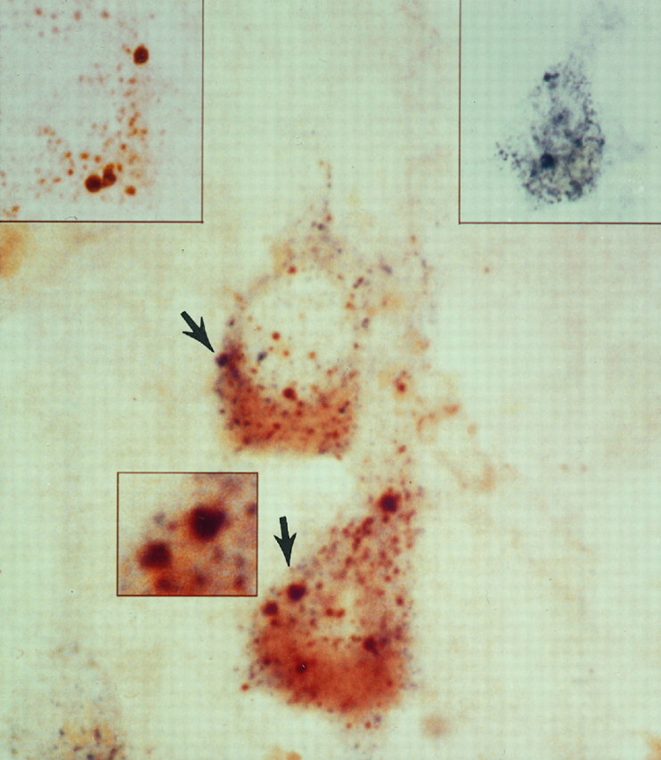
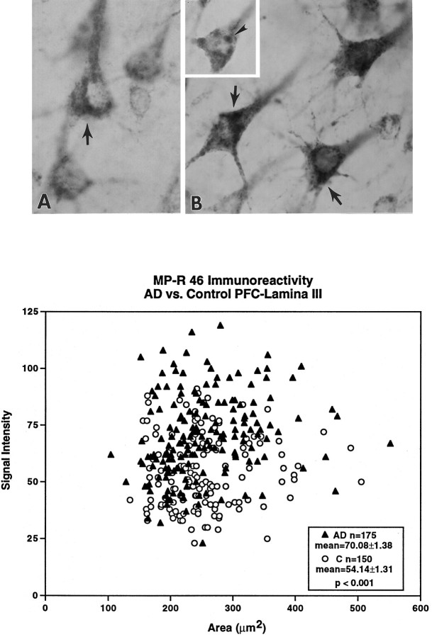
The early endosome is the first vacuolar compartment along the endocytic pathway. It is the site of internalization and initial processing of amyloid precursor protein (APP) and apolipoprotein E (ApoE), two proteins of etiological importance in Alzheimer's disease, and a putative site of beta-amyloid peptide (Abeta) formation. Here, we identify early endosomes in human pyramidal neurons, using specific compartmental markers and morphometry, and show that in Alzheimer's disease individual endosomes display up to 32-fold larger volumes than the normal average. Endosomal enlargement contributed to an average 2.5-fold larger total endosomal volume per neuron, implying a marked increase in endocytic activity. Endosomal alterations were evident in most pyramidal neurons in Alzheimer brain, detectable at early stages of the disease but absent in several other neurodegenerative disorders examined. In addition, mature and proenzyme forms of the proteases cathepsin B and cathepsin D, a candidate APP secretase, were identified in most early endosomes in Alzheimer brains but were detectable in only a minor proportion of endosomes in normal brain. Expression of the cation-dependent 46 kDa mannose 6-phosphate receptor was elevated in pyramidal neurons of Alzheimer brains, which could be a possible basis for the altered cathepsin trafficking pattern. Enhanced endocytic activity, coupled with increased trafficking to endosomes of proteases, which may have the ability under pathological conditions to generate Abeta, constitutes a potential mechanism by which beta-amyloidogenesis may become accelerated in sporadic AD and also be subject to influences by ApoE.


Figures

Fig. 1.
Fig. 1.
Identification of early endosomes as distinct hydrolase-containing compartments in human pyramidal neurons.A, Consistent with established morphological criteria, rab5-positive early endosomes (arrowhead) in cortical pyramids of aged control human brain were distributed close to the plasmalemma and were relatively uniform in size. Immunoreactive endosomes were located principally in the soma and proximal dendrites. We have shown that these structures represent a subpopulation of neuronal acid–hydrolase-containing compartments (see Fig. 3).B, We distinguished early endosomal compartments from a smaller subgroup of hydrolase-containing, rab5-negative, MP-R215-positive late endosomes (arrowhead) that displayed the typical perinuclear distribution. C, Lysosomes, which contain Cat D (arrow) and a number of other hydrolases, are MP-R-negative, rab5-negative, and distinct from early and late endosomes. Lysosomes, which comprise the major population of acidic vacuolar compartments, are typically 50–400 nm in diameter and are distributed uniformly throughout the cytosol. Magnification in A–C, 4500×).
Fig. 2.
Fig. 2.
Morphometric analysis of rab5-positive early endosomes in control and Alzheimer’s disease (AD) brains. High-magnification images of early endosomal compartments (arrows) in neocortical neurons in lamina III of aged control (A) and AD (B) brains immunostained with rab5 and enhanced by Nomarski optics show the striking increase in the size of these compartments in AD brain, as compared with control. In some, invaginations suggest a location at the plasmalemma. C, Percentage of cell area occupied by rab5-positive early endosomes plotted as a function of total cell area. Alterations in early endosomes were widespread in AD brains, and many pyramidal neurons in AD brains (filled triangles) exhibited a significant increase (>2 SD higher than the control mean) in total endosomal volume per cross-sectional cell area as compared with age-matched controls (open circles) (AD mean = 5.37% ± 0.19; control mean = 2.18% ± 0.09). D, The size distributions of individual early endosomes present in 25 lamina III pyramids from each of eight aged control and nine AD brains show that a substantial proportion of individual neuronal endosomes in the AD brains had volumes larger than those in control brains. Endosomal volume is expressed as a function of cell area for easier visualization of data. E, The percentage of abnormally large endosomal profiles that are >1 μm3. Values are mean percentage based on 25 pyramidal neurons per individual brain (n = 9 AD and 8 controls). Approximately 10-fold higher numbers are seen in AD brains as compared with controls (AD mean = 7.8% ± 1.65; control mean = 0.80% ± 0.13). Changes in endosomal volume as a function of cell area were not significant in AD and control brains. Magnification inA, B, 4700×.
Fig. 3.
Fig. 3.
Colocalization of mature proteases in early endosomal compartments of human neurons. Immunofluorescence confocal images of a neocortical pyramidal neuron from control (A–C) and AD (D–F) brains, using antisera to mature Cat B (red) and rab5 (green), demonstrate that mature lysosomal hydrolases often reside in early endosomes (yellow). The localization of mature protease in early endosomal compartments occurred to a greater extent in neurons in the AD brains than in controls and more often in the larger endosomal profiles. Both Cat B and Cat D (not shown) were present in rab5-positive early endosomes as well as rab5-negative late endosomes, lysosomes, and lipofuscin. Magnification in A–F, 4000×.
Fig. 4.
Fig. 4.
Immature proteases frequently are detected in early endosomes of AD neurons. Double-label immunocytochemical analysis with bright-field microscopy and antisera directed against pro-Cat D (brown, left inset) and rab5 (blue, right inset) in this lamina III pyramid from an AD brain is a representative example of the frequent colocalization of immature proteases in the abnormally large early endosomes (arrows). Pro-Cat D is also present in a population of smaller immunoreactive vacuolar compartments, which are consistent with late endosomes and are increased in number in AD neurons, as compared with normal controls (see Fig.1B). Magnification, 4350×; magnification inleft and right insets, 3600×; magnification in center inset, 6000×.
Fig. 5.
Fig. 5.
Levels of MP-R46 are elevated in neurons of AD brains. Immunocytochemical studies using antibody directed against MP-R46 show increased levels of this receptor (arrows) in lamina III neurons in the AD brains (B) than in controls (A). (The cells in Arange in optical density from 57 to 67, control mean = 54.14 ± 1.31; those in B range in optical density from 71 to 86, AD mean = 70.80 ± 1.38; p < 0.001. Percentage difference above the mean optical density was equivalent for neurons inA vs B.) In AD brains displaying lower levels of immunostaining, MP-R46 immunolabeling could be visualized clearly within large vacuolar profiles (B, inset, arrowhead) that resembled the abnormally large rab5-positive early endosomal profiles. Semiquantitative morphometric analysis from sections of the prefrontal cortex revealed that cortical pyramids in laminae III of the AD brains ( filled triangles) exhibit increased MP-R46 densities per cross-sectional area as compared with age-matched controls (open circles). Magnification inA, B, B inset, 3600×.

References

    1. Araki W, Kunishita T, Takahashi K, Ikeda S, Tabina T. Demonstration of amyloid β-protein secretion in a mouse neuronal cell line. Neurosci Lett. 1994;167:125–127. - PubMed
    1. Berg T, Gjoen T, Bakke O. Physiological functions of endosomal proteolysis. Biochem J. 1995;307:313–326. - PMC - PubMed
    1. Bernstein HG, Wiedanders B. An immunohistochemical study of cathepsin E in Alzheimer-type dementia brains. Brain Res. 1994;667:287–290. - PubMed
    1. Bleekemolen JE, Stein M, von Figura K, Slot JW, Geuze HJ. The two mannose 6-phosphate receptors have almost identical subcellular distributions in U937 monocytes. Eur J Cell Biol. 1988;47:366–372. - PubMed
    1. Brown WJ, Goodhouse J, Farquhar MG. Mannose 6-phosphate receptors for lysosomal enzymes cycle between the Golgi complex and endosomes. J Cell Biol. 1985;103:1235–1247. - PMC - PubMed

Publication types

MeSH terms