. 1997 Aug 15;17(16):6203-12.
doi: 10.1523/JNEUROSCI.17-16-06203.1997.

Growth factor activity of endothelin-1 in primary astrocytes mediated by adhesion-dependent and -independent pathways

Affiliations

Growth factor activity of endothelin-1 in primary astrocytes mediated by adhesion-dependent and -independent pathways

S Cazaubon et al. J Neurosci. .

Abstract

Endothelin-1 (ET-1) has been shown to induce DNA synthesis in primary astrocytes by stimulating the extracellular signal-regulated kinase (ERK) pathway. To clarify the mechanisms responsible for the anchorage-dependent growth of astrocytes, the relationships between cell adhesion and ERK activation were investigated. Here it is reported that ET-1 promotes the formation of stress fibers and focal adhesions and the tyrosine phosphorylation of focal adhesion kinase (FAK) and paxillin, as well as Src activation and association of phosphorylated FAK with Grb2. Pretreatment of astrocytes with cytochalasin D or C3-transferase, which inhibits actin polymerization or Rho activity, respectively, prevented the activation/phosphorylation of Src, FAK, and paxillin after ET-1 stimulation; by contrast, the ERK pathway was not significantly affected. This differential activation of FAK/Src and ERK pathways was also observed with astrocytes 10 and 60 min after replating on poly-L-ornithine-precoated dishes. Collectively, these findings indicate that activation of FAK and Src is dependent on actin cytoskeleton integrity, Rho activation, and adhesion to extracellular matrix, whereas ERK activation is independent of these intracellular events and seems to correlate with activation of the newly identified protein tyrosine kinase PYK2. Induction of DNA synthesis by ET-1, however, was reduced dramatically in astrocytes pretreated with either cytochalasin D or C3-transferase. This study provides a demonstration of Rho- and adhesion-dependent activation of FAK/Src, which collaborates with adhesion-independent activation of PYK2/ERK for DNA synthesis in ET-1-stimulated astrocytes.


Figures

Fig. 1.
Fig. 1.
Confocal analysis of the cytoskeleton organization in ET-1-treated astrocytes. Quiescent astrocytes (top row) or astrocytes treated with 50 nm ET-1 for 10 min (bottom row) were labeled using FITC-conjugated phalloidin (green), anti-vinculin antibodies, or anti-phosphotyrosine antibodies (PY20) plus anti-mouse antibodies conjugated to Cy3 (red). The results are representative of five independent experiments.
Fig. 2.
Fig. 2.
Tyrosine phosphorylation or autophosphorylation of immunoprecipitated FAK, paxillin, and Src in ET-1-treated astrocytes. Lysates from untreated quiescent astrocytes (C) and those treated for 10 min with 50 nm ET-1 (ET) or AlF4 (30 mmsodium fluoride and 10 μm aluminum chloride) (AlF) were immunoprecipitated with anti-FAK (1 μg), anti-paxillin (anti-Pax) (2.5 μg), or anti-Src (2.5 μg) antibodies, respectively. Immunoprecipitated proteins were analyzed either by immunoblotting with anti-phosphotyrosine antibodies [WB anti-Y(P): IP anti-FAK, IP anti-Pax] or by their ability to undergo autophosphorylation [Auto(P): IP anti-Src]. After stripping of the bound antibodies, the same membrane was reincubated with anti-FAK, anti-Pax, and anti-Src antibodies, respectively, showing that a comparable amount of proteins was immunoprecipitated. Molecular mass markers (kDa) are shown on the left side. Arrowheadsindicate the heavy chain of immunoglobulins (H), and arrows indicate the migration of the immunoprecipitated proteins FAK, Pax, or Src. The results are representative of three independent experiments.
Fig. 3.
Fig. 3.
Association of tyrosine-phosphorylated FAK and Shc to the SH2 domain of Grb2 in ET-1-treated astrocytes. A, Quiescent astrocytes were not treated (C) or were treated for 10 min with either 50 nm ET-1 (ET) or AlF4 (30 mmsodium fluoride and 10 μm aluminum chloride) (AlF), and the ability of SOS, Shc, and FAK to interact with either the GST-Grb2 or GST-Grb3–3 fusion proteins, immobilized on glutathione-agarose beads, was determined by immunoblotting with anti-SOS, anti-Shc, or anti-FAK antibodies, respectively. After stripping of the bound antibodies, the membrane was reincubated with anti-phosphotyrosine antibodies [WB anti-Y(P)]. B, Lysates from astrocytes untreated (0), treated for 10 min with ET-1 at the indicated concentrations (2, 10, 50 nm) (left), or treated with 50 nm ET-1 for the indicated times (5, 10, 20, 40, 60 min) (right) were precipitated with GST-Grb2. Bound proteins were analyzed by immunoblotting with either anti-Shc or anti-FAK antibodies. The results are representative of three independent experiments.
Fig. 4.
Fig. 4.
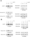
Effect of cytochalasin D and C3-transferase pretreatment on the ET-1 responses of astrocytes. Quiescent astrocytes were pretreated either with 2 μm cytochalasin D for 2 hr (A) or with 1.5 μg/ml of C3 for 24 hr (B) before addition of 50 nm ET-1 for 10 min (ET); untreated cells (C). Cell lysates were then submitted to precipitation with GST-Grb2 fusion protein plus immunoblotting with either anti-SOS (1 μg/ml) or anti-Shc antibodies (1 μg/ml) (GST-Grb2); immunoblot analysis using anti-Raf-1 (1 μg/ml) and anti-ERK2 antibodies (0.5 μg/ml) (cell lysates); immunoprecipitation with anti-FAK (1 μg), anti-paxillin (anti-Pax) (2.5 μg), or anti-Src (2.5 μg) antibodies, respectively. Immunoprecipitated proteins were analyzed either by immunoblotting with anti-phosphotyrosine antibodies [WB anti-Y(P), IP anti-FAK, IP anti-Pax] or by their ability to undergo autophosphorylation [auto(P): IP anti-Src]. The results are representative of four independent experiments.
Fig. 5.
Fig. 5.
Tyrosine phosphorylation of immunoprecipitated PYK2 in ET-1- and AlF4-treated astrocytes. Astrocytes were not pretreated (−) or were pretreated for 18 hr with 0.1 μg/mlPTX or 160 nmTPA for 24 hr with 1.5 μg/ml C3-transferase (C3), or for 2 hr with 2 μm cytochalasin D (CytoD) in serum-free medium before addition of effectors for 10 min: 50 nm ET-1 (ET), AlF4 (30 mmsodium fluoride and 10 μm aluminum chloride) (AlF), or 160 nmTPA; untreated cells (C). Cell lysates were then submitted either to immunoblot analysis using anti-ERK2 antibodies (0.5 μg/ml) (cell lysates) or to immunoprecipitation with anti-PYK2 antibodies (10 μl). Immunoprecipitated proteins were analyzed by immunoblotting with anti-phosphotyrosine antibodies [WB anti-Y(P)]. The results are representative of three independent experiments.
Fig. 6.
Fig. 6.
Effect of matrix attachment on the ET-1 responses of astrocytes. Quiescent serum-starved astrocytes were replated on precoated poly-l-ornithine glass coverslips or dishes of 60 mm diameter. Cells were allowed to attach 10 min, 60 min, or 15 hr before treatment with 50 nm ET-1 for 10 min (ET); untreated cells (C).A, Confocal analysis of the cytoskeleton organization using FITC-conjugated phalloidin (green).B, Cell lysates were submitted either to immunoblot analysis using anti-ERK2 antibodies (0.5 μg/ml) (cell lysates) or to immunoprecipitation with anti-Src (2.5 μg), anti-FAK (1 μg), anti-paxillin (anti-Pax) (2.5 μg), or anti-PYK2 antibodies (10 μl), respectively. Immunoprecipitated proteins were analyzed either by their ability to undergo autophosphorylation [Auto(P)] or by immunoblotting with anti-phosphotyrosine antibodies [WB anti-Y(P)]. The results are representative of four independent experiments.
Fig. 6.
Fig. 6.
Effect of matrix attachment on the ET-1 responses of astrocytes. Quiescent serum-starved astrocytes were replated on precoated poly-l-ornithine glass coverslips or dishes of 60 mm diameter. Cells were allowed to attach 10 min, 60 min, or 15 hr before treatment with 50 nm ET-1 for 10 min (ET); untreated cells (C).A, Confocal analysis of the cytoskeleton organization using FITC-conjugated phalloidin (green).B, Cell lysates were submitted either to immunoblot analysis using anti-ERK2 antibodies (0.5 μg/ml) (cell lysates) or to immunoprecipitation with anti-Src (2.5 μg), anti-FAK (1 μg), anti-paxillin (anti-Pax) (2.5 μg), or anti-PYK2 antibodies (10 μl), respectively. Immunoprecipitated proteins were analyzed either by their ability to undergo autophosphorylation [Auto(P)] or by immunoblotting with anti-phosphotyrosine antibodies [WB anti-Y(P)]. The results are representative of four independent experiments.
Fig. 7.
Fig. 7.
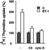
Effect of cytochalasin D and C3-transferase pretreatment on [3H]thymidine uptake in ET-1-treated astrocytes. Quiescent astrocytes in 96-well plates were not pretreated (−) or were pretreated either with 1.5 μg/ml of C3-transferase (C3) or 2 μm cytochalasin D for 2 hr (cyto D) before incubation with 50 nm ET-1 (ET-1) for 24 hr; untreated cells (C). [3H]thymidine (1 μCi/well) was added 3 hr before the cells were harvested. Data are means of 12 determinations ± SEM and expressed as percentages of [3H]thymidine uptake of untreated cells (280 ± 18). The results are of one experiment representative of three.
Fig. 8.
Fig. 8.
Schematic representation of adhesion-dependent and -independent pathways leading to DNA synthesis in ET-1-treated astrocytes. Primary astrocytes express ET-1-receptors (ETB-R subtype) coupled via PTX-insensitive heterotrimeric G-proteins (Gq) to at least two distinct pathways: (1) the adhesion-independent activation of the ERK pathway (gray arrows) initiated by phosphorylation of Shc by a protein tyrosine kinase (PTK); PYK2 activation coincides with ERK activation and could be responsible for this phosphorylation event; and (2) the adhesion-dependent activation of FAK/Src pathway (black arrows), which requires Rho activation and stress fiber formation. Adhesion of astrocytes to ECM proteins involves integrins, heterodimeric receptors composed of α and β chains. Both signaling pathways cooperate to induce DNA synthesis in response to ET-1.

Similar articles

Cited by

References

    1. Cazaubon SM, Couraud PO. Nitric oxide and endothelin at the blood-brain barrier. An introduction to the blood-brain barrier Pardridge WM. 1997. Cambridge UP; Cambridge, in press.
    1. Cazaubon SM, Ramos-Morales F, Fischer S, Schweighoffer F, Strosberg AD, Couraud PO. Endothelin induces tyrosine phosphorylation and GRB2 association of SHC in astrocytes. J Biol Chem. 1994;269:24805–24809. - PubMed
    1. Cazaubon S, Parker PJ, Strosberg AD, Couraud PO. Endothelins stimulate phosphorylation and activity of p42/mitogen-activated protein kinase in astrocytes. Biochem J. 1993;293:381–386. - PMC - PubMed
    1. Cobb BS, Schaller MD, Leu TH, Parsons JT. Stable association of pp60src and pp59fyn with the focal adhesion-associated protein tyrosine kinase, pp125FAK. Mol Cell Biol. 1994;14:147–155. - PMC - PubMed
    1. Couraud PO, Durieu-Trautmann O, Le Nguyen P, Marin P, Glibert F, Strosberg AD. Functional endothelin-1 receptors in rat astrocytoma C6. Eur J Pharmacol. 1991;206:191–198. - PubMed

Publication types

MeSH terms

Substances