. 1997 Aug 15;17(16):6213-25.
doi: 10.1523/JNEUROSCI.17-16-06213.1997.

Lack of neurotrophin 3 causes losses of both classes of spiral ganglion neurons in the cochlea in a region-specific fashion

Affiliations

Lack of neurotrophin 3 causes losses of both classes of spiral ganglion neurons in the cochlea in a region-specific fashion

B Fritzsch et al. J Neurosci. .

Abstract

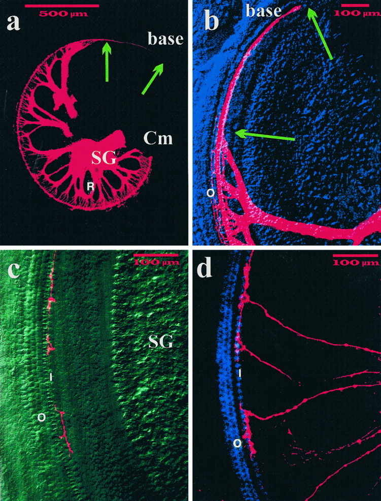
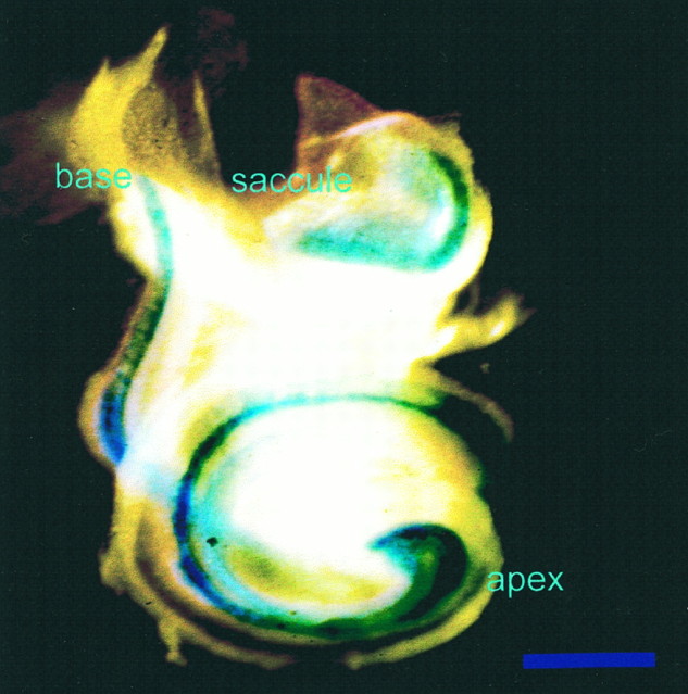
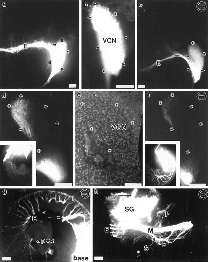
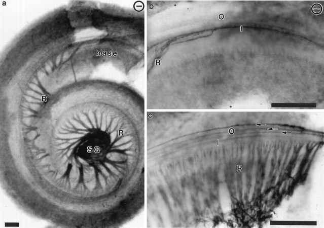
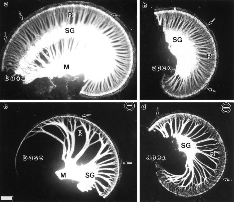
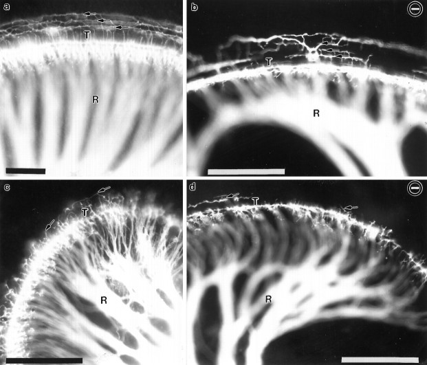
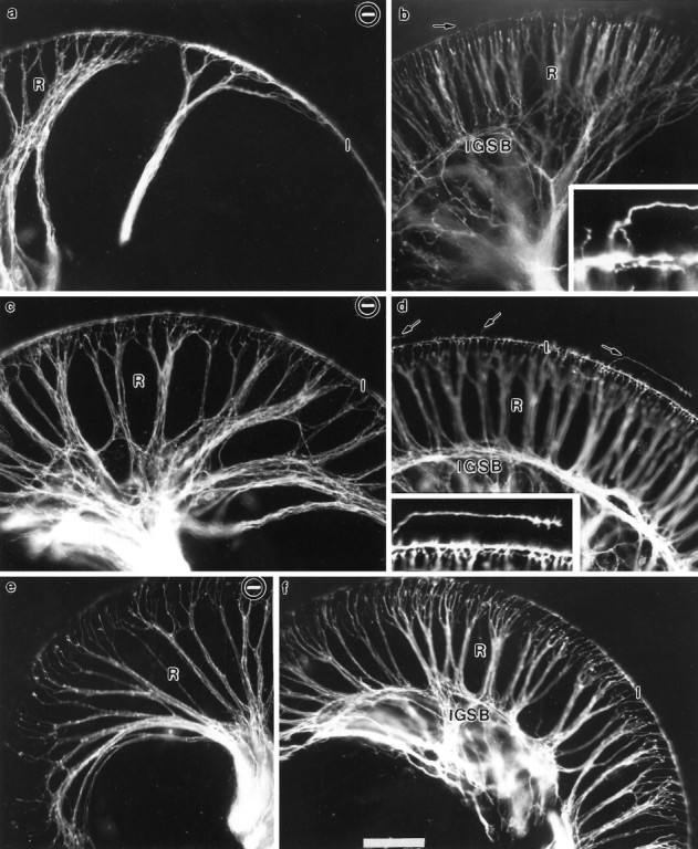
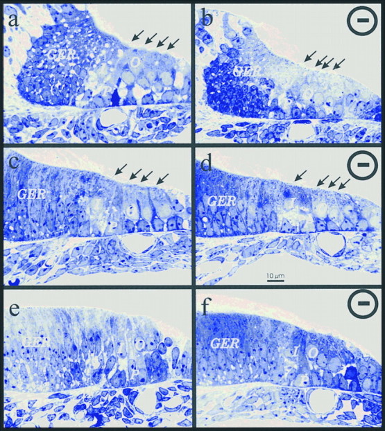
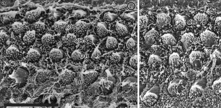
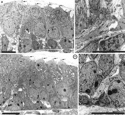
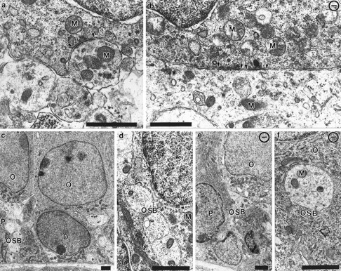
Essential functions of neurotrophin 3 (NT-3) in regulating afferent and efferent innervation of the cochlea have been characterized by comparison of normal and NT-3 mutant mice. NT-3 deficiency has striking, region-specific effects, with complete loss of sensory neurons in the basal turn and dramatic but incomplete neuronal loss in the middle and apical turns. The sensory innervation of inner and outer hair cells was reorganized in mutant animals. Instead of a strictly radial pattern of innervation, the axons of remaining sensory neurons projected spirally along the row of inner hair cells to innervate even the most basal inner hair cells. Innervation of outer hair cells was strongly reduced overall and was not detected in the basal turn. The presence of fibers extending to both inner and outer hair cells suggests that subsets of types I and II sensory neurons survive in the absence of NT-3. Likewise, projections of the cochlea to auditory nuclei of the brainstem were attenuated but otherwise present. Equally striking changes in efferent innervation were observed in mutant animals that closely mimicked the abnormal sensory innervation pattern. Despite these impressive innervation deficiencies, the morphology of the organ of Corti and the development of inner and outer hair cells appeared comparatively normal.


Figures

Fig. 4.
Fig. 4.
Distribution of sensory afferent fibers in the basal turn of P0 NT-3 mutant homozygotes. All afferents (a, b) and subsets of afferents (c, d) were filled with DiI from the brainstem. Note a that the basal turn of the cochlea (base) lacks spiral ganglion cells (SG) and radial fibers (R), both of which are present in the middle turn (Cm). Nevertheless, fibers extend along inner hair cells toward the base (arrows). As illustrated b, few fibers extend to outer hair cells (O) near the middle turn. Selective labeling reveals the distribution of individual afferents in the middle basal turn (c, d). Note that the terminal arbor of each afferent fiber is restricted to several inner hair cells (I) with no processes crossing to the three rows of outer hair cells (O), which appear normally developed in this differential interference contrast (c) or fluorescence image (d). Scale bar, 500 μm for all images.
Fig. 2.
Fig. 2.
Distribution of β-galactosidase in the whole-mounted cochlea of a newborn mouse heterozygous for the targeted replacement of the NT-3 coding exon with a construct containing alacZ gene cDNA. Reporter expression is an indicator of the endogenous pattern of NT-3 expression. Reaction product is visible throughout the cochlea. The reaction is most prominent in the apex but also is prominent in the middle and basal turns. Reaction product is visible in the saccular sensory epithelium, but no labeling is apparent in the spiral ganglion. Scale bar, 1 mm.
Fig. 6.
Fig. 6.
Comparison of afferent projections to the ventral cochlear nucleus in wild-type (a, b, d, e) and mutant (c, f) mice and the application site in the ear (g, h). Coronal sections of brainstem labeled from the cochlea are shown (a–f). Labeled cochlear afferent fibers extend throughout the ventral cochlear nucleus (VCN) in both wild-type (a, b) and NT-3 mutant (c) mice near the eighth nerve root. Note the apparent reduction in number of efferents (E ina, c) in the mutant as well as a less dense labeling near the dorsolateral aspect of the cochlear nucleus in the mutant. The cochlear projection covers the ventral cochlear nucleus (asterisks; compare b, e, images at two different excitations from the same section) as much in the control (a) as in the NT-3 mutant littermate (c). A selective projection from the basal turn (d, f, insets) shows a comparable pattern of projection to the dorsomedial aspect of the ventral cochlear nucleus (shown in the Hoechst-stained control by asterisks ine). However, there is a reduction in fiber density in the mutant that compares with the reduced density of innervation of the basal turn of the cochlea. Closer examination of the cochlea and the spiral ganglion with the modiolus in an NT-3 mutant with a basal turn application (g, h) shows numerous efferent collaterals (E) extending throughout the cochlea (g). In addition, spiral ganglion cells near the middle turn are labeled, as are efferent and afferent fibers in the modiolus (M) and toward the brain (h). Note that no ganglion neurons are labeled near the modiolus, where efferents (E) diverge within the inferior vestibular ganglion toward the posterior vertical canal (P) and the saccule (S). The arrow indicates the same fibers from the middle turn; E indicates the same efferent fascicles from the apex (g, h). Scale bars, 100 μm.
Fig. 1.
Fig. 1.
Patterns of cochlear innervation in whole mounts of newborn wild-type and NT-3 mutant homozygotes. An mAb to acetylated tubulin was used to reveal the nerve fiber patterns in a whole-mounted cochlea of a newborn NT-3 mutant (a), in the basal turn of a newborn NT-3 mutant (b), and in a control littermate (c). In the cochlea of the NT-3 mutant, spiral ganglion cells (SG) are present in the middle turn and extend their fibers to the apical turn. No radial fibers (R) exist near the basal turn. However, fibers extend from the middle turn spiral ganglion cells along the inner hair cells of the base (b) but do not extend to the layer of outer hair cells. In contrast, in the control littermate a dense radial fiber innervation is seen near the base (c), which extends along inner hair cells (I) and all three rows (arrows) of outer hair cells (O). ⊖, NT-3 mutant in this and all following figures. Scale bars, 100 μm.
Fig. 3.
Fig. 3.
DiI-labeling patterns of afferent innervation of the cochlea in newborn wild-type and NT-3-deficient mice. These images show differences in the pattern of innervation between a control (a, b) and an NT-3-deficient littermate (c, d). DiI was implanted in the eighth nerve afferents in the brainstem and diffused for 4.5 d at 37°C. The cochlea was split into a basal (a, c) and an apical (b, d) half. Note the change of course of radial fibers (R) in the NT-3 mutant littermate near the base (a) and the apex (b). There is a complete absence of the spiral ganglia (SG) in the basal half turn (c) and some reduction near the apex (d). However, fibers extend along the inner hair cells to the very tip of the base. M, Modiolus. Scale bar, 100 μm for all images.
Fig. 5.
Fig. 5.
Comparison of afferent innervation to the middle and apical turns in newborn wild-type and NT-3 mutant mice. The distribution of afferents to the middle turn (a, b) and the apex (c, d) is shown for a control (a, c) and an NT-3 deficient littermate (b, d). Note the numerous fibers that extend beyond the inner hair cells and through the tunnel of Corti (T) to the three rows of outer hair cells in normal animals (arrows ina, c). Fibers to outer hair cells are reduced in number, and their distribution is more patchy in the NT-3 null mice in both the middle (arrows in b) and the apical turn (arrows in d). R, Fibers. Scale bars, 100 μm.
Fig. 7.
Fig. 7.
Comparisons of distributions of olivo-cochlear efferent fibers in newborn wild-type and NT-3 mutant mice. The distribution of olivo-cochlear efferent fibers was revealed by implanting DiI into the floor of the fourth ventricle where these fibers cross. a, c, e, Innervation in NT-3−/− mouse;b, d, f, innervation in corresponding regions of a control littermate. The distribution is shown for the base (a, b), middle turn (c, d) and apex (e, f) of the cochlea. Note that the distribution of efferent fibers follows closely the distribution of afferents and shows very similar differences between the control and NT-3−/− mice. In control mice, some efferents extend to outer hair cells (d, arrows and inset). This occurs only rarely in NT-3 mutant mice (not shown). In control mice there is an elaborate network of efferent fibers within the spiral ganglion and a clear formation of IGSBs, which cannot be identified in the NT-3 mutant mice. The overall progression of efferent fibers to the apex is comparable in both normal and mutant mice (e, f), but again fewer fibers are seen in the NT-3-deficient mice (e).I, Inner spiral bundle near the inner hair cells. Scale bar, 100 μm for all images.
Fig. 8.
Fig. 8.
Differentiation of the sensory epithelium of the cochlea in normal and NT-3-deficient mice. These micrographs of 1-μm-thick plastic sections taken from newborn mice show the appearance of inner (I) and outer (O) hair cells in the basal (a, b), middle (c, d), and apical turn (e, f) of a control (a, c, e) and an NT-3 null littermate (b, d, f). Note that the only difference that is readily apparent is the height of the greater epithelial ridge (compare a, b), a transitory structure that degenerates in neonates. The only other difference that is apparent is the almost complete absence of nerve fibers entering the organ of Corti through the habenula perforata (H). Note that the position of the pilar cells (P) is always next to the lateral wall of the spiral vessel underneath the basilar membrane.Arrows indicate the apical specializations protruding into the scala media. Scale bar, 10 μm for all panels.
Fig. 9.
Fig. 9.
Differentiation of inner and outer hair cells in newborn control and NT-3 mutant mice. Scanning electron micrographs from the apical (a) and middle turn (b) of an NT-3 mutant mouse (a) and a control littermate (b). Note that a single row of inner hair cells (IH) and multiple rows of outer hair cells (OH) are present in both preparations. The appearance of the hair cells, each of which has developed an array of apical stereocilia and a single kinocilium (arrows), appears to be the same in the normal and mutant animals. Scale bar, 10 μm for both images.
Fig. 10.
Fig. 10.
Transmission electron microscopic images of the organ of Corti in newborn control and mutant animals. Electron micrographs show the organ of Corti with the three rows of outer hair cells (O), Deiter’s cells (D), one row of inner hair cells (I), and pilar cells (P) in control (a) and NT-3 mutant (c) animals. Other than the plane of the section, which is slightly oblique in c, there are no differences between the control (a) and the NT-3 mutant littermate (c). Note the presence of apical specializations in all hair cells (arrows in a, c). The habenula perforata (H) has no fibers passing through at the basal turn of NT-3 mutant mice (c, d) but has numerous fibers joining the inner spiral bundle (ISB) underneath the inner hair cell in control animals (a, b). Scale bar, 10 μm in each panel.
Fig. 11.
Fig. 11.
Electron microscopic examination of contact development in control and NT-3-deficient animals. These transmission electron micrographs show the synaptic interactions with the inner hair cells (a, b) and approach of fibers to outer hair cells (c–f) in a control (a, c, d) and an NT-3 mutant mouse (b, e, f). Inner hair cells show synaptic contact regions, which occasionally bear presynaptic bars surrounded by synaptic vesicles with adjacent fiber profiles (arrows in a, b). In contrast, the contacts are not as mature on outer hair cells and do not show clear indications of synaptic specializations. In control mice many fibers running in the outer spiral bundles (OSB) are seen underneath every hair cell (c, d). In the mutants, few fibers are restricted to the innermost outer hair cell next to the pilar cells (e), and only a few fibers are next to the outer hair cells (f, O). P, Pilar cells; D, Deiter’s cells; M, mitochondria. Scale bar, 1 μm in each panel.

Similar articles

Cited by

References

    1. Berglund AM, Benson TE, Brown MC. Synapses from labeled type II axons in the mouse cochlear nucleus. Hear Res. 1996;94:31–46. - PubMed
    1. Bianchi LM, Cohan CS. Effects of the neurotrophins and CNTF on developing statoacoustic neurons: comparison with an otocyst-derived factor. Dev Biol. 1993;159:353–365. - PubMed
    1. Bianchi LM, Conover JC, Fritzsch B, De Chiara T, Lindsay RM, Yancopoulos GD. Degeneration of vestibular neurons in late embryogenesis of both heterozygous and homozygous BDNF null mutant mice. Development. 1996;122:1965–1973. - PubMed
    1. Easter SS, Ross LS, Frankfurter A. Initial tract formation in the mouse brain. J Neurosci. 1993;13:285–299. - PMC - PubMed
    1. Echteler SM. Developmental segregation in the afferent projections to mammalian auditory hair cells. Proc Natl Acad Sci USA. 1992;89:6324–6327. - PMC - PubMed

Publication types

MeSH terms

LinkOut - more resources