. 1997 Aug 15;17(16):6325-37.
doi: 10.1523/JNEUROSCI.17-16-06325.1997.

P2 receptor excitation of rodent hypoglossal motoneuron activity in vitro and in vivo: a molecular physiological analysis

Affiliations

P2 receptor excitation of rodent hypoglossal motoneuron activity in vitro and in vivo: a molecular physiological analysis

G D Funk et al. J Neurosci. .

Abstract

The role of P2 receptors in controlling hypoglossal motoneuron (XII MN) output was examined (1) electrophysiologically, via application of ATP to the hypoglossal nucleus of rhythmically active mouse medullary slices and anesthetized adult rats; (2) immunohistochemically, using an antiserum against the P2X2 receptor subunit; and (3) using PCR to identify expression of P2X2 receptor subunits in micropunches of tissue taken from the XII motor nucleus. Application of ATP to the hypoglossal nucleus of mouse medullary slices and anesthetized rats produced a suramin-sensitive excitation of hypoglossal nerve activity. Additional in vitro effects included potentiation of inspiratory hypoglossal nerve output via a suramin- and pyridoxal-phosphate-6-azophenyl-2',4'-disulphonic acid (PPADS)-sensitive mechanism, XII MN depolarization via activation of a suramin-sensitive inward current, decreased neuronal input resistance, and a slow-onset theophylline-sensitive reduction of inspiratory output likely resulting from hydrolysis of extracellular ATP to adenosine and activation of P1 receptors. Immunohistochemically, P2X2 receptors were detected in inspiratory XII MNs that were labeled with Lucifer yellow. These data, combined with identification of mRNA for three P2X2 receptor subunit isoforms within the hypoglossal nucleus (two of which have not been localized previously in brain) and the previous demonstration that P2X receptors are ubiquitously expressed in cranial and spinal motoneuron pools, support not only a role of P2 receptors in modulating inspiratory hypoglossal activity but a general role of P2 receptors in modulating motor outflow from the CNS.


Figures

Fig. 6.
Fig. 6.
Excitation of XII nerve activity in vivo. A, Tonic excitation of the ipsilateral XII nerve activity after 7.6 sec application of 10 mm ATP into the left XII nucleus under control conditions (i), 10 min after a 7.6 sec application of 10 mm suramin (ii), and 30 min after suramin application (iii). Traces represent integrated activity from the left and right XII nerve (LXII,RXII), phrenic nerve (Phr), and tracheal pressure (TP; calibration, 0–10 cm H2O). B, Application of 10 mmATP into the rostral portion of the XII motor nucleus (1 mm rostral to obex) potentiated inspiratory burst amplitude. (Note that burst amplitude potentiation was observed in only 2 of 13 trials.)Traces represent integrated activity from the left and right XII nerve (LXII, RXII), arterial blood pressure (BP; calibration, 100–200 mm Hg), and raw phrenic nerve activity (Phr).C, Antidromic field potentials generated by stimulation of the ipsilateral XII nerve and recorded within the hypoglossal motor nucleus were used to indicate pipette placement (i).ii, An example of inspiratory-related extracellular action potentials (Unit) to verify ATP injection within the vicinity of inspiratory-modulated XII MNs. D, Expanded time scale recording showing the discharge envelope of inspiratory activity recorded bilaterally from XII nerves and unilaterally from the phrenic nerve (end-tidal CO2, 5.5%).
Fig. 1.
Fig. 1.
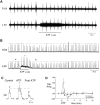
Excitation of XII nerve activity in vitro by ATP. A, Bilateral recordings of XII nerve activity of a medullary slice from a postnatal day 2 mouse showing the effects of 30 sec, 1 mm ATP injected unilaterally over the ventromedial portion of the LXII nucleus. Rhythmic bursts of activity represent inspiratory-related XII nerve activity. B, Rectified and integrated signal of the raw data traces and ATP response shown in A illustrates the ATP-mediated potentiation of burst amplitude and subsequent decrease in burst amplitude. Note the slower time scale in B.C, The individual control (#), ATP (*), and post-ATP (§) bursts indicated in B are shown in expanded form. The baseline shift present during ATP application has been removed to facilitate comparison of burst amplitude. D, Time course of the changes in XII nerve inspiratory amplitude after local application of 1 mm ATP to the ventromedial portion of the XII motor nucleus (n = 20). Drug application occurred at time = 0. Effects on contralateral XII nerve output are documented also; asterisk indicates significant difference from control levels.
Fig. 2.
Fig. 2.
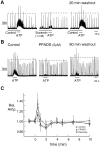
Antagonism of ATP excitatory responses by suramin and PPADS. A, The tonic excitatory and amplitude-potentiating effects produced by ATP were reduced significantly when ATP was applied for the last 10 sec of a 30 sec suramin (1 mm) application, although the post-ATP inhibition was not blocked (middle panel). Inhibitory action of suramin was removed after 20 min of washout (right panel). B, Bath application of PPADS caused a significant reduction in the excitatory effects of ATP without affecting the post-ATP decrease in inspiratory burst amplitude (middle panel). The PPADS block of the inspiratory burst amplitude-potentiating component of the ATP response was reversible, given sufficient recovery time, whereas the tonic excitatory effect did not show complete recovery even after 90 min of washout. C, Time course of the changes in XII nerve inspiratory burst amplitude after local application of ATP (1 mm) before, during, and after bath application of PPADS (n = 5). ATP application occurred at time =0; asterisk indicates significant difference from amplitude values observed at the same time during the control ATP application.
Fig. 3.
Fig. 3.
ATP-mediated inward currents in inspiratory XII MNs. A, A 30 sec application of 1 mm ATP over XII motor nucleus produced tonic excitation of the nerve, increased XII nerve inspiratory burst amplitude (top trace, ∫XII), and induced a 75 pA inward current in an inspiratory XII MN. Voltage ramps (−100 mV to −45 mV conducted over 2 sec) performed during control and ATP application (indicated byasterisk) are plotted versus current. The ATP-induced current, obtained by subtracting the control from the ATP curve (ATP-Control), shows weak inward rectification.B, Inward current induced by 10 sec of 1 mmATP is blocked by 30 sec preapplication of 1 mm suramin.C, Inward current produced by 15 sec application of 10 mm ATP over inspiratory XII MN after bath application of TTX. The current responses to 10 mV hyperpolarizing voltage steps during control (Control) and during a subsequent ATP application (ATP) indicated a decrease in input resistance (right panel).
Fig. 4.
Fig. 4.
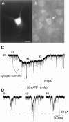
Lucifer yellow-labeled inspiratory MN (A) shows immunofluorescence for P2X2R (B, arrow). The neuron labeled in A and B responded to local application of 1 mm ATP with a 75 pA inward current and a decrease in input resistance (C). Inspiratory synaptic currents recorded during control (#1), early during ATP application (#2), and late in the ATP application (#3) are shown with an expanded time scale in D. Holding potential, −60 mV; peaks markedsynaptic currents represent 3 of 11 inspiratory synaptic currents; asterisk equals −10 mV pulses for calculating neuronal input resistance. Scale bar in A (applies toB), 25 μm.
Fig. 7.
Fig. 7.
P2X2R immunostaining in the neonatal mouse and adult rat XII nucleus. A–C, Transverse sections through postnatal day 2 neonatal mouse medulla ∼100 μm rostral to obex, showing P2X2R TRITC immunofluorescence (1:100 P2X296ab) at increasing magnification. Thewhite box outline in B is the region shown in C to illustrate greater cytoplasmic versus nuclear staining. D, E, Transverse sections through adult rat medulla ∼100 μm caudal to obex, showing P2X2 receptor immunoperoxidase staining (1:2000 P2X296ab) of XII MNs. F, G, Transverse sections through adult rat medulla immediately caudal to obex, showing P2X2 receptor immunoperoxidase staining of XII MNs under test conditions (1:4000 P2X2R96ab) (F) and block of the immunolabeling by antiserum preadsorbed with its target peptide (G). Scale bars in A–G represent 100, 50, 10, 100, 20, 150, and 150 μm, respectively.dmx, Dorsal motor nucleus of vagus; XII, hypoglossal motor nucleus (arrows inA).
Fig. 5.
Fig. 5.
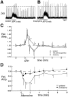
Post-ATP inhibition of inspiratory burst amplitude results from activation of A1 adenosine receptors.A, Inhibition of inspiratory burst amplitude after 30 sec application of ATP is blocked (B) by 90 sec preapplication of theophylline (100 μm).C, Time course of the changes in XII nerve inspiratory burst amplitude after local application of ATP (1 mm) before, during, and after local application of theophylline (100 μm; n = 8); asteriskindicates significant difference from amplitude values observed at the same time during the control ATP application. D, Time course of the changes in XII nerve inspiratory burst amplitude after local application of adenosine (1 mm; n= 6); asterisk indicates significant difference from control, pre-ATP levels.
Fig. 8.
Fig. 8.
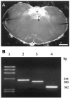
Identification of mRNA for P2X2receptor subunit isoforms in micropunches from the hypoglossal nucleus.A, Dark-field photomicrograph of 240 μm transverse medullary section showing location of micropunch (arrowhead). B, Ethidium bromide-stained gel showing the PCR products for three P2X2 receptor subunit isoforms isolated from the micropunch. Lane 1, Molecular weight marker (φX174/HaeIII; BRL, Bethesda, MD). Lane 2, The 85 bp insert isoform (Housley et al., 1995) obtained by using primers 1558as/S4. Lane 3, Isoform (Brake et al., 1994) obtained by using primers 1558as/S3.Lane 4, Isoform (Brändle et al., 1997) with 207 bp exon deletion obtained by using primers 1558as/S1. Lengths of fragments are indicated on the right. Calibration bar inA, 1 mm.

Similar articles

Cited by

References

    1. Abbracchio MP, Burnstock G. Purinoceptors: are there families of P2X and P2Y purinoceptors? Pharmacol Ther. 1994;64:445–475. - PubMed
    1. Aldes LD, Chapman ME, Chronister RB, Haycock JW. Sources of noradrenergic afferents to the hypoglossal nucleus of the rat. Brain Res Bull. 1992;29:931–942. - PubMed
    1. Akasu T, Hirai K, Koketsu K. Modulatory actions of ATP on membrane potentials of bullfrog sympathetic ganglion cells. Brain Res. 1983;258:313–317. - PubMed
    1. Balcar VJ, Li Y, Killinger S, Bennett MR. Autoradiography of P2X ATP receptors in the rat brain. Br J Pharmacol. 1995;115:302–306. - PMC - PubMed
    1. Bellingham MC, Berger AJ. Adenosine suppresses excitatory glutamatergic inputs to rat hypoglossal motoneurons in vitro. Neurosci Lett. 1994;177:143–146. - PubMed

Publication types

MeSH terms

LinkOut - more resources