

Massive autaptic self-innervation of GABAergic neurons in cat visual cortex
- PMID: 9236244
- PMCID: PMC6568358
- DOI: 10.1523/JNEUROSCI.17-16-06352.1997
Massive autaptic self-innervation of GABAergic neurons in cat visual cortex
Abstract
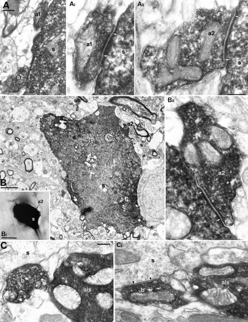
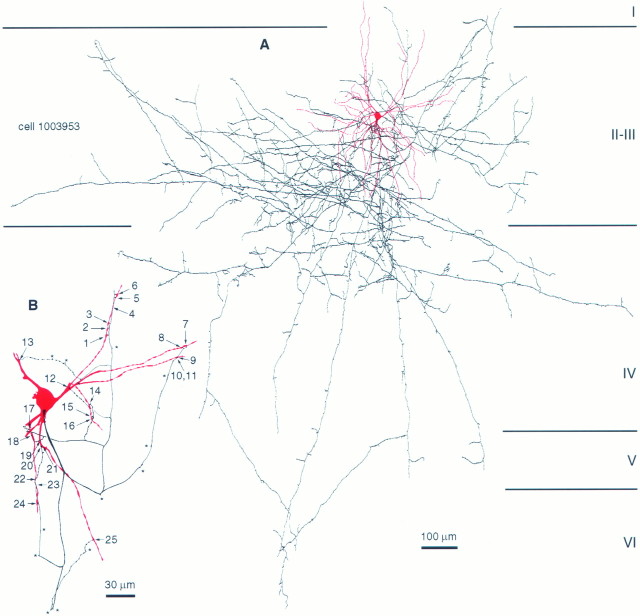
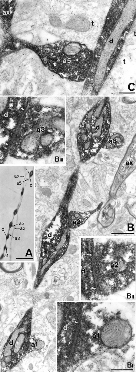
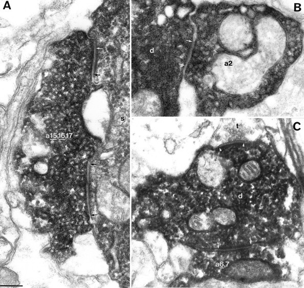
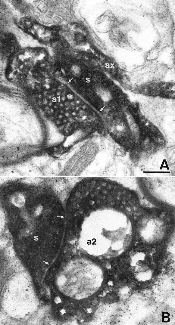
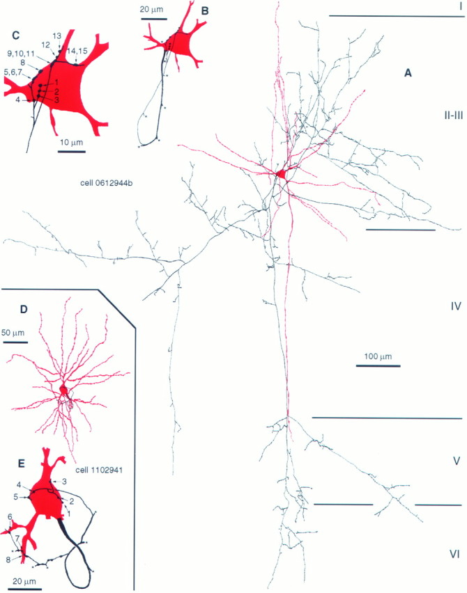
Autapses are transmitter release sites made by the axon of a neuron on its own dendrites. We determined the numbers and precise subcellular position of autapses on different spiny and smooth dendritic cell types using intracellular biocytin filling in slices of adult neocortex. Potential self-innervation was light microscopically assessed on 10 pyramidal cells, 7 spiny stellate cells, and 41 smooth dendritic neurons from cortical layers II-V. Putative autapses occurred on each smooth dendritic neuron and on seven pyramids, but not on spiny stellate cells. However, electron microscopic examination of all light microscopically predicted sites on pyramids (n = 28) showed only one case of self-innervation with two autapses on dendritic spines. Interneurons were classified by postsynaptic target distribution () and all putative autapses of seven basket, three dendrite-targeting, and three double bouquet cells were scrutinized. All basket and dendrite-targeting cells established self-innervation, the number of autapses being 12 +/- 7 and 22 +/- 12 (mean +/- SD), respectively; only one of the double bouquet cells formed autapses (n = 3). Basket cell autapses (n = 74) were closer to the soma (12.2 +/- 22.3 microm) than autapses established by dendrite-targeting cells (51.8 +/- 49.9 microm; n = 66). The degree of self-innervation is cell type-specific. Unlike on spiny cells, autapses are abundant on GABAergic basket and dendrite-targeting interneurons, with subcellular location similar to that of synapses formed by the parent cell on other neurons. The extensive self-innervation may modulate integrative properties and/or the firing rhythm of the neuron in a manner temporally correlated with its own activity.
Figures


Similar articles
-
Fast IPSPs elicited via multiple synaptic release sites by different types of GABAergic neurone in the cat visual cortex.J Physiol. 1997 May 1;500 ( Pt 3)(Pt 3):715-38. doi: 10.1113/jphysiol.1997.sp022054. J Physiol. 1997. PMID: 9161987 Free PMC article.
-
Differentially interconnected networks of GABAergic interneurons in the visual cortex of the cat.J Neurosci. 1998 Jun 1;18(11):4255-70. doi: 10.1523/JNEUROSCI.18-11-04255.1998. J Neurosci. 1998. PMID: 9592103 Free PMC article.
-
Effect, number and location of synapses made by single pyramidal cells onto aspiny interneurones of cat visual cortex.J Physiol. 1997 May 1;500 ( Pt 3)(Pt 3):689-713. doi: 10.1113/jphysiol.1997.sp022053. J Physiol. 1997. PMID: 9161986 Free PMC article.
-
[Cytophysiology of spiny stellate cells of striate cortex and their role in excitatory mechanisms of intracortical synaptic circulation].Morfologiia. 2005;128(5):7-19. Morfologiia. 2005. PMID: 16669238 Review. Russian.
-
GABAergic inhibition in the neostriatum.Prog Brain Res. 2007;160:91-110. doi: 10.1016/S0079-6123(06)60006-X. Prog Brain Res. 2007. PMID: 17499110 Review.
Cited by
-
The Diversity of Cortical Inhibitory Synapses.Front Neural Circuits. 2016 Apr 25;10:27. doi: 10.3389/fncir.2016.00027. eCollection 2016. Front Neural Circuits. 2016. PMID: 27199670 Free PMC article. Review.
-
Physiological features of parvalbumin-expressing GABAergic interneurons contributing to high-frequency oscillations in the cerebral cortex.Curr Res Neurobiol. 2023 Dec 16;6:100121. doi: 10.1016/j.crneur.2023.100121. eCollection 2024. Curr Res Neurobiol. 2023. PMID: 38616956 Free PMC article. Review.
-
Combined two-photon imaging, electrophysiological, and anatomical investigation of the human neocortex in vitro.Neurophotonics. 2014 Jul;1(1):011013. doi: 10.1117/1.NPh.1.1.011013. Epub 2014 Sep 11. Neurophotonics. 2014. PMID: 26157969 Free PMC article.
-
Wiring Specificity and Synaptic Diversity in the Mouse Lateral Central Amygdala.J Neurosci. 2016 Apr 20;36(16):4549-63. doi: 10.1523/JNEUROSCI.3309-15.2016. J Neurosci. 2016. PMID: 27098697 Free PMC article.
-
Microcircuits and their interactions in epilepsy: is the focus out of focus?Nat Neurosci. 2015 Mar;18(3):351-9. doi: 10.1038/nn.3950. Nat Neurosci. 2015. PMID: 25710837 Free PMC article. Review.
References
-
- Aghajanian GK, Rasmussen K. Intracellular studies in the facial nucleus illustrating a simple new method for obtaining viable motoneurons in adult rat brain slices. Synapse. 1989;3:331–338. - PubMed
-
- Anderson JC, Douglas RJ, Martin KAC, Nelson JC. Map of the synapses formed with the dendrites of spiny stellate neurons of cat visual cortex. J Comp Neurol. 1994;341:25–38. - PubMed
-
- Beaulieu C, Somogyi P. Targets and quantitative distribution of GABAergic synapses in the visual cortex of the cat. Eur J Neurosci. 1990;2:296–303. - PubMed
Publication types
MeSH terms
Substances
Grants and funding
LinkOut - more resources
Full Text Sources
Miscellaneous